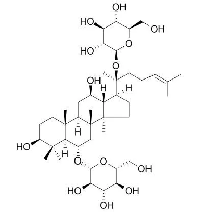
| Description: |
Ginsenoside Rg1 has antiaging, anti-platelet activation, promotion of wound healing, and neuroprotective effects, it has protective effect against Aβ25-35-induced toxicity in PC12 cells,might be through the insulin-like growth factor-I receptor (IGF-IR) and estrogen receptor (ER)signaling pathways. Ginsenoside Rg1 is a desirable agent for enhancing CD4+ T-cell activity, as well as the correction of Th1-dominant pathological disorders, which by increasing Th2 specific cytokine secretion and by repressing Th1 specific cytokine production. It increased the expression of the vascular endothelial growth factor (VEGF) mRNA and reduced ERK pathway, expression of transforming growth factor beta (TGF-β) mRNA. |
| In vitro: |
| Acta Pharmacol Sin. 2009 Mar;30(3):299-306. | | Ginsenoside Rg1 promotes endothelial progenitor cell migration and proliferation.[Pubmed: 19262553] | To investigate the effect of Ginsenoside Rg1 on the migration, adhesion, proliferation, and VEGF expression of endothelial progenitor cells (EPCs).
METHODS AND RESULTS:
EPCs were isolated from human peripheral blood and incubated with different concentrations of Ginsenoside Rg1 (0.1, 0.5, 1.0, and 5.0 micromol/L) and vehicle controls. EPC migration was detected with a modified Boyden chamber assay. EPC adhesion was determined by counting adherent cells on fibronectin-coated culture dishes. EPC proliferation was analyzed with the 3-(4,5-dimethylthiazol-2-yl)-2,5-diphenyltetrazolium bromide (MTT) assay. In vitro vasculogenesis was assayed using an in vitro vasculogenesis detection kit. A VEGF-ELISA kit was used to measure the amount of VEGF protein in the cell culture medium. Ginsenoside Rg1 promoted EPC adhesion, proliferation, migration and in vitro vasculogenesis in a dose- and time-dependent manner. Cell cycle analysis showed that 5.0 micromol/L of Ginsenoside Rg1 significantly increased the EPC proliferative phase (S phase) and decreased the resting phase (G(0)/G(1) phase). Ginsenoside Rg1 increased vascular endothelial growth factor production.
CONCLUSIONS:
The results indicate that Ginsenoside Rg1 promotes proliferation, migration, adhesion and in vitro. | | Int Immunopharmacol. 2004 Feb;4(2):235-44. | | Ginsenoside Rg1 enhances CD4(+) T-cell activities and modulates Th1/Th2 differentiation.[Pubmed: 14996415] | Panax ginseng is commonly used as a tonic medicine in Asian countries such as Korea, China, and Japan. It has been reported that Ginsenoside Rg1 in P. ginseng increases the proportion of T helper (Th) cells among the total number of T cells and promotes IL-2 gene expression in murine splenocytes. This implies that Ginsenoside Rg1 increases the immune activity of CD4(+) T cells, however, the exact mechanism remains unknown.
METHODS AND RESULTS:
The present study elucidated the direct effect of Rg1 on helper T-cell activities and on Th1/Th2 lineage development. The results demonstrated that Ginsenoside Rg1 had no mitogenic effects on unstimulated CD4(+) T cells, but augmented CD4(+) T-cell proliferation upon activation with anti-CD3/anti-CD28 antibodies in a dose-dependent manner. Rg1 also enhanced the expression of cell surface protein CD69 on CD4(+) T cells. In Th0 condition, Ginsenoside Rg1 increases the expression of IL-2 mRNA, and enhances the expression of IL-4 mRNA on CD4(+) T cells, suggesting that Rg1 prefers to induce Th2 lineage development. In addition, Ginsenoside Rg1 increases IL-4 secretion in CD4(+) T cells under Th2 skewed condition, while decreasing IFN-gamma secretion of cells in Th1 polarizing condition.
Thus, Rg1 enhances Th2 lineage development from the naïve CD4(+) T cell both by increasing Th2 specific cytokine secretion and by repressing Th1 specific cytokine production.
CONCLUSIONS:
Therefore, these results suggest that Ginsenoside Rg1 is a desirable agent for enhancing CD4(+) T-cell activity, as well as the correction of Th1-dominant pathological disorders. | | Oncotarget . 2017 Jul 24;8(33):55384-55393. | | Ginsenoside Rg1 attenuates adjuvant-induced arthritis in rats via modulation of PPAR-γ/NF-κB signal pathway[Pubmed: 28903427] | | Ginsenoside Rg1, the main active compound in Panax ginseng, has already been shown to have anti-inflammatory effects. However, the protective effects of Rg1 on rheumatoid arthritis (RA) remain unclear. The aim of the present study was to investigate the effects and mechanisms of Rg1 on adjuvant-induced arthritis (AIA) in rats. AIA rats were given Rg1 at doses of 5, 10, and 20 mg/kg intraperitoneally for 14 days to observe the anti-arthritic effects. The results showed that Rg1 significantly alleviated joint swelling and injuries. Rg1 can also significantly reduce the level of TNF-α and IL-6, increase PPAR-γ protein expression, inhibit IκBα phosphorylation and NF-κB nuclear translocation in the inflammatory joints of AIA rats and RAW264.7 cells stimulated by lipopolysaccharide (LPS). The results indicate that Rg1 has therapeutic effects on AIA rats, and the mechanism might be associated with its anti-inflammatory effects by up-regulating PPAR-γ and subsequent inhibition of NF-κB signal pathway. | | J Ginseng Res . 2016 Jan;40(1):9-17. | | A UPLC/MS-based metabolomics investigation of the protective effect of ginsenosides Rg1 and Rg2 in mice with Alzheimer's disease[Pubmed: 26843817] | | Background: Alzheimer's disease (AD) is a progressive brain disease, for which there is no effective drug therapy at present. Ginsenoside Rg1 (G-Rg1) and G-Rg2 have been reported to alleviate memory deterioration. However, the mechanism of their anti-AD effect has not yet been clearly elucidated.
Methods: Ultra performance liquid chromatography tandem MS (UPLC/MS)-based metabolomics was used to identify metabolites that are differentially expressed in the brains of AD mice with or without ginsenoside treatment. The cognitive function of mice and pathological changes in the brain were also assessed using the Morris water maze (MWM) and immunohistochemistry, respectively.
Results: The impaired cognitive function and increased hippocampal Aβ deposition in AD mice were ameliorated by G-Rg1 and G-Rg2. In addition, a total of 11 potential biomarkers that are associated with the metabolism of lysophosphatidylcholines (LPCs), hypoxanthine, and sphingolipids were identified in the brains of AD mice and their levels were partly restored after treatment with G-Rg1 and G-Rg2. G-Rg1 and G-Rg2 treatment influenced the levels of hypoxanthine, dihydrosphingosine, hexadecasphinganine, LPC C 16:0, and LPC C 18:0 in AD mice. Additionally, G-Rg1 treatment also influenced the levels of phytosphingosine, LPC C 13:0, LPC C 15:0, LPC C 18:1, and LPC C 18:3 in AD mice.
Conclusion: These results indicate that the improvements in cognitive function and morphological changes produced by G-Rg1 and G-Rg2 treatment are caused by regulation of related brain metabolic pathways. This will extend our understanding of the mechanisms involved in the effects of G-Rg1 and G-Rg2 on AD. |
|
| In vivo: |
| J Gerontol A Biol Sci Med Sci. 2014 Mar;69(3):282-94. | | Long-term ginsenoside Rg1 supplementation improves age-related cognitive decline by promoting synaptic plasticity associated protein expression in C57BL/6J mice.[Pubmed: 23833204 ] | In aging individuals, age-related cognitive decline is the most common cause of memory impairment. Among the remedies, Ginsenoside Rg1, a major active component of ginseng, is often recommended for its antiaging effects. However, its role in improving cognitive decline during normal aging remains unknown and its molecular mechanism partially understood.
METHODS AND RESULTS:
This study employed a scheme of Rg1 supplementation for female C57BL/6J mice, which started at the age of 12 months and ended at 24 months, to investigate the effects of Rg1 supplementation on the cognitive performance. We found that Rg1 supplementation improved the performance of aged mice in behavior test and significantly upregulated the expression of synaptic plasticity-associated proteins in hippocampus, including synaptophysin, N-methyl-D-aspartate receptor subunit 1, postsynaptic density-95, and calcium/calmodulin-dependent protein kinase II alpha, via promoting mammalian target of rapamycin pathway activation.
CONCLUSIONS:
These data provide further support for Rg1 treatment of cognitive degeneration during aging. | | Thromb Res. 2014 Jan;133(1):57-65. | | Ginsenoside Rg1 inhibits platelet activation and arterial thrombosis.[Pubmed: 24196231 ] | Derived from the root of Panax ginseng C.A.Mey, Panax notoginsenosides (PNS) is a widely used herbal medicine to treat atherothrombotic diseases in Asian medicine. Ginsenoside Rg1 is one of the main compounds responsible for the pharmaceutical actions of PNS. As platelets play pivotal roles in atherothrombogenesis, we therefore studied the effect of Rg1 on platelet activation and its underlying mechanisms.
METHODS AND RESULTS:
Human platelets are obtained from healthy subjects. Platelet activation and the inhibition of Rg1 were assessed by Born aggregometer, flow cytmetry, flow chamber and western blot. The in vivo thrombosis model was induced by 10% FeCl3 on mesenteric arterioles of wild type B57/b6 mice.
Rg1 significantly inhibited platelet aggregation induced by thrombin, ADP, collagen and U46619, e.g., aggregation rate stimulated by 0.1UmL(-1) thrombin was decreased 46% by Rg1. Rg1 also reduced thrombin (0.1UmL(-1))-enhanced fibrinogen binding and P-selectin expression of single platelet by 81% and 66%, respectively. Rg1 affected αIIbβ3-mediated outside-in signaling as demonstrated by diminished platelet spreading on immobilized fibrinogen. Rg1 also decreased the rate of clot retraction in platelet rich plasma. Furthermore, Rg1 decreased platelet adhesion on collagen surface under a shear rate correlated to the arterial flow (1000s(-1)) by approximately 70%. Western blot showed that Rg1 potently inhibited ERK phosphrylation. The in vitro findings were further evaluated in the mouse model of in vivo arterial thrombosis, and Rg1 was found to prolong the mesenteric arterial occlusion time (34.9±4.1min without and 64.3±4.9min with Rg1; p<0.01).
CONCLUSIONS:
Rg1 inhibits platelet activation via the inhibition of ERK pathway, and attenuates arterial thrombus formation in vivo. |
|


 Cell. 2018 Jan 11;172(1-2):249-261.e12. doi: 10.1016/j.cell.2017.12.019.IF=36.216(2019)
 Cell Metab. 2020 Mar 3;31(3):534-548.e5. doi: 10.1016/j.cmet.2020.01.002.IF=22.415(2019)
 Mol Cell. 2017 Nov 16;68(4):673-685.e6. doi: 10.1016/j.molcel.2017.10.022.IF=14.548(2019)