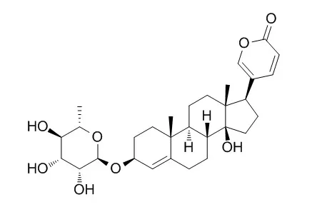
| Kinase Assay: |
| Oxidative Medicine & Cellular Longevity, 2018, 2018:1-17. | | Proscillaridin A Promotes Oxidative Stress and ER Stress, Inhibits STAT3 Activation, and Induces Apoptosis in A549 Lung Adenocarcinoma Cells.[Reference: WebLink] | Cardiac glycosides are natural compounds used for the treatment of cardiovascular disorders. Although originally prescribed for cardiovascular diseases, more recently, they have been rediscovered for their potential use in the treatment of cancer. Proscillaridin A (PSD-A), a cardiac glycoside component of Urginea maritima, has been reported to exhibit anticancer activity. However, the cellular targets and anticancer mechanism of PSD-A in various cancers including lung cancer remain largely unexplored.
METHODS AND RESULTS:
In the present study, we found that PSD-A inhibits growth and induces apoptosis in A549 lung adenocarcinoma cells. The anticancer activity of PSD-A was found to be associated with the activation of JNK, induction of ER stress, mitochondrial dysfunction, and inhibition of STAT3 activation. PSD-A induces oxidative stress as evidenced from ROS generation, GSH depletion, and decreased activity of TrxR1. PSD-A-mediated ER stress was verified by increased phosphorylation of eIF2α and expression of its downstream effector proteins ATF4, CHOP, and caspases-4. PSD-A triggered apoptosis by inducing JNK (1/2) activation, increasing bax/bcl-2 ratio, dissipating mitochondrial membrane potential, and inducing cleavage of caspases and PARP. Further study revealed that PSD-A inhibits both constitutive and inducible STAT3 activations and decreases STAT3 DNA-binding activity. Moreover, PSD-A-mediated inhibition of STAT3 activation was found to be associated with increased SHP-1 expression, decreased phosphorylation of Src, and binding of PSD-A with STAT3 SH2 domain. Finally, STAT3 knockdown by shRNA inhibited growth and enhanced apoptotic efficacy of PSD-A.
CONCLUSIONS:
Taken together, the data suggest that PSD-A could be developed into a potential therapeutic agent against lung adenocarcinoma. | | Cell Death & Disease,2018,9(10):984. | | Proscillaridin A exerts anti-tumor effects through GSK3β activation and alteration of microtubule dynamics in glioblastoma.[Reference: WebLink] | Glioblastoma (GBM) is characterized by highly aggressive growth and invasive behavior. Due to the highly lethal nature of GBM, new therapies are urgently needed and repositioning of existing drugs is a promising approach.
METHODS AND RESULTS:
We have previously shown the activity of Proscillaridin A (ProA), a cardiac glycoside inhibitor of the Na(+)/K(+) ATPase (NKA) pump, against proliferation and migration of GBM cell lines. ProA inhibited tumor growth in vivo and increased mice survival after orthotopic grafting of GBM cells. This study aims to decipher the mechanism of action of ProA in GBM tumor and stem-like cells. ProA displayed cytotoxic activity on tumor and stem-like cells grown in 2D and 3D culture, but not on healthy cells as astrocytes or oligodendrocytes. Even at sub-cytotoxic concentration, ProA impaired cell migration and disturbed EB1 accumulation at microtubule (MT) plus-ends and MT dynamics instability. ProA activates GSK3β downstream of NKA inhibition, leading to EB1 phosphorylation on S155 and T166, EB1 comet length shortening and MT dynamics alteration, and finally inhibition of cell migration and cytotoxicity. Similar results were observed with digoxin.
CONCLUSIONS:
Therefore, we disclosed here a novel pathway by which ProA and digoxin modulate MT-governed functions in GBM tumor and stem-like cells. Altogether, our results support ProA and digoxin as potent candidates for drug repositioning in GBM. |
|
| Cell Research: |
| Archives of Pharmacal Research, 2007, 30(10):1216-1224. | | Apoptosis-mediated cytotoxicity of ouabain, digoxin and proscillaridin A in the estrogen independent MDA-MB-231 breast cancer cells.[Reference: WebLink] |
METHODS AND RESULTS:
We examined the effects of three cardiac glycosides, ouabain, digoxin and Proscillaridin A, on the proliferation of estrogen independent MDA-MB-231 breast cancer cells. In terms of reduction in cell viability, the compounds rank for both 24 h and 48 h of incubation in MDA-MB-231 cells in the order Proscillaridin A > digoxin > ouabain. Digoxin for 24 h and 48 h of incubation in MDA-MB-231 cells proved to be only slightly more potent than ouabain, with IC50 values of 122 +/- 2 and 70 +/- 2 nM, respectively, compared to 150 +/- 2 and 90 +/- 2 nM for ouabain. In contrast, Proscillaridin A, was much more active and showed a high level of cytotoxic potency, IC50 51 +/- 2 and 15 +/- 2 nM for 24 h and 48 h of incubation, respectively. The concentrations of digoxin, ouabain and Proscillaridin A needed to inhibit [3H]thymidine incorporation into DNA by 50% (IC50) in MDA-MB-231 cells for 24 h of incubation were found to be 124 +/- 2 nM, 142 +/- 2 nM, and 48 +/- 2 nM, respectively.
CONCLUSIONS:
In the present study, we demonstrated that ouabain, digoxin, and Proscillaridin A induce apoptosis in MDA-MB-231 cells by increasing free calcium concentration and by activating caspase-3. |
|


 Cell. 2018 Jan 11;172(1-2):249-261.e12. doi: 10.1016/j.cell.2017.12.019.IF=36.216(2019)
 Cell Metab. 2020 Mar 3;31(3):534-548.e5. doi: 10.1016/j.cmet.2020.01.002.IF=22.415(2019)
 Mol Cell. 2017 Nov 16;68(4):673-685.e6. doi: 10.1016/j.molcel.2017.10.022.IF=14.548(2019)