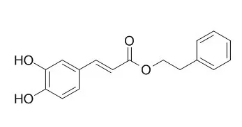
| In vitro: |
| Immunopharmacol Immunotoxicol. 2014 Oct;36(5):371-7. | | Caffeic acid phenethyl ester inhibits the inflammatory effects of interleukin-1β in human corneal fibroblasts.[Pubmed: 25151996] | The regulatory mechanisms of Caffeic acid phenethyl ester on cellular signaling pathways were examined using Western blot and electrophoretic mobility shift assays.
METHODS AND RESULTS:
A functional validation was carried out by evaluating the inhibitory effects of Caffeic acid phenethyl ester on neutrophil and monocyte migration in vitro. Caffeic acid phenethyl ester inhibited the expression of IL-6, MCP-1 and ICAM-1 induced by the pro-inflammatory cytokine IL-1β in corneal fibroblasts. The activation of AKT and NF-κB by IL-1β was markedly inhibited by Caffeic acid phenethyl ester, whereas the activity of mitogen-activated protein kinases (MAPKs) was not affected. Caffeic acid phenethyl ester significantly suppressed the IL-1β-induced migration of differentiated (d)HL-60 and THP-1 cells.
CONCLUSIONS:
These anti-inflammatory effects of Caffeic acid phenethyl ester may be expected to inhibit the infiltration of leukocytes into the corneal stroma in vivo. | | Oncotarget . 2015 Mar 30;6(9):6684-707. | | Caffeic acid phenethyl ester induced cell cycle arrest and growth inhibition in androgen-independent prostate cancer cells via regulation of Skp2, p53, p21Cip1 and p27Kip1[Pubmed: 25788262] | | Abstract
Prostate cancer (PCa) patients receiving the androgen ablation therapy ultimately develop recurrent castration-resistant prostate cancer (CRPC) within 1-3 years. Treatment with Caffeic acid phenethyl ester (CAPE) suppressed cell survival and proliferation via induction of G1 or G2/M cell cycle arrest in LNCaP 104-R1, DU-145, 22Rv1, and C4-2 CRPC cells. CAPE treatment also inhibited soft agar colony formation and retarded nude mice xenograft growth of LNCaP 104-R1 cells. We identified that CAPE treatment significantly reduced protein abundance of Skp2, Cdk2, Cdk4, Cdk7, Rb, phospho-Rb S807/811, cyclin A, cyclin D1, cyclin H, E2F1, c-Myc, SGK, phospho-p70S6kinase T421/S424, phospho-mTOR Ser2481, phospho-GSK3α Ser21, but induced p21Cip1, p27Kip1, ATF4, cyclin E, p53, TRIB3, phospho-p53 (Ser6, Ser33, Ser46, Ser392), phospho-p38 MAPK Thr180/Tyr182, Chk1, Chk2, phospho-ATM S1981, phospho-ATR S428, and phospho-p90RSK Ser380. CAPE treatment decreased Skp2 and Akt1 protein expression in LNCaP 104-R1 tumors as compared to control group. Overexpression of Skp2, or siRNA knockdown of p21Cip1, p27Kip1, or p53 blocked suppressive effect of CAPE treatment. Co-treatment of CAPE with PI3K inhibitor LY294002 or Bcl-2 inhibitor ABT737 showed synergistic suppressive effects. Our finding suggested that CAPE treatment induced cell cycle arrest and growth inhibition in CRPC cells via regulation of Skp2, p53, p21Cip1, and p27Kip1.
Keywords: Skp2; Caffeic acid phenethyl ester; cell cycle arrest; p53; prostate cancer. |
|
| In vivo: |
| Am J Otolaryngol. 2014 Jul-Aug;35(4):482-6. | | Effects of caffeic acid phenethyl ester on wound healing of nasal mucosa in the rat: an experimental study.[Pubmed: 24767474] | In this experimental study, our aim was to use histopathological examination to investigate the effects of Caffeic acid phenethyl ester on the wound healing of rat nasal mucosa after mechanical trauma.
METHODS AND RESULTS:
The rats were randomly divided into 3 experimental groups: a non-treated group (n=7), a control saline group (n=7) and a Caffeic acid phenethyl ester group (n=7). The non-treated group received no treatment for 15 days. The second group was administered saline (2.5 mL/kg, intraperitoneal) once a day for 15 days. The third group received Caffeic acid phenethyl ester intraperitoneally at a dose of 10 μmol/kg once a day for 15 days. At the beginning of the study, unilateral mechanical nasal trauma was induced on the right nasal mucosa of all rats in the three groups using a brushing technique. Samples were stained using hematoxylin and eosin solution and were examined by a pathologist using a light microscope. The severity of inflammation was milder in the Caffeic acid phenethyl ester group compared with that in the non-treated and saline groups (P<0.05). The subepithelial thickness index was lower in the experimental group (P<0.05). Goblet cell and ciliated cell loss was substantially reduced in the experimental group compared with the non-treated and saline groups (P<0.05).
CONCLUSIONS:
Caffeic acid phenethyl ester decreases inflammation and the loss of goblet cells and ciliated cells. Therefore, Caffeic acid phenethyl ester has potential beneficial effects on the wound healing of nasal mucosa in the rat. | | Epilepsy Behav. 2013 Nov;29(2):275-80. | | Caffeic acid phenethyl ester prevents apoptotic cell death in the developing rat brain after pentylenetetrazole-induced status epilepticus.[Pubmed: 24012504] | The aim of this study was to investigate whether Caffeic acid phenethyl ester exerts neuroprotective effects on the developing rat brain after status epilepticus.
METHODS AND RESULTS:
Twenty-one dams reared Wistar male rats, and 21-day-old rats were divided into three groups: control group, pentylenetetrazole-induced status epilepticus group, and Caffeic acid phenethyl ester-treated group. Status epilepticus was induced on the first day of experiment. Caffeic acid phenethyl ester injections (30 mg/kg intraperitoneally) started 40 min after the tonic phase of status epilepticus was reached, and the injections of Caffeic acid phenethyl ester were repeated over 5 days. Rats were sacrificed, and brain tissues were collected on the 5th day of experiment after the last injection of Caffeic acid phenethyl ester. Apoptotic cell death was evaluated. Histopathological examination showed that Caffeic acid phenethyl ester significantly preserved the number of neurons in the CA1, CA3, and dentate gyrus regions of the hippocampus and the prefrontal cortex. It also diminished apoptosis in the hippocampus and the prefrontal cortex.
CONCLUSIONS:
In conclusion, this experimental study suggests that Caffeic acid phenethyl ester administration may be neuroprotective in status epilepticus in the developing rat brain. |
|


 Cell. 2018 Jan 11;172(1-2):249-261.e12. doi: 10.1016/j.cell.2017.12.019.IF=36.216(2019)
 Cell Metab. 2020 Mar 3;31(3):534-548.e5. doi: 10.1016/j.cmet.2020.01.002.IF=22.415(2019)
 Mol Cell. 2017 Nov 16;68(4):673-685.e6. doi: 10.1016/j.molcel.2017.10.022.IF=14.548(2019)