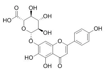
| Kinase Assay: |
| J Ethnopharmacol. 2015 Mar 13;162:69-78. | | Biological evaluation and molecular docking of baicalin and scutellarin as Helicobacter pylori urease inhibitors.[Pubmed: 25557028] |
METHODS AND RESULTS:
Baicalin and Scutellarin effectively suppressed Helicobacter pylori urease in dose-dependent and time-independent manner with IC50 of 0.82±0.07 mM and 0.47±0.04 mM, respectively, compared to AHA (IC50=0.14±0.05 mM). Structure-activity relationship disclosed 4'-hydroxyl gave flavones an advantage to binding with Helicobacter pylori urease. Kinetic analysis revealed that the types of inhibition were non-competitive and reversible with inhibition constant Ki of 0.14±0.01 mM and 0.18±0.02 mM for baicalin and Scutellarin, respectively. The mechanism of urease inhibition was considered to be blockage of the SH groups of Helicobacter pylori urease, since thiol reagents (L,D-dithiothreitol, L-cysteine and glutathione) abolished the inhibitory action and competitive active site Ni(2+) binding inhibitors (boric acid and sodium fluoride) carried invalid effect. Molecular docking study further supported the structure-activity analysis and indicated that baicalin and Scutellarin interacted with the key residues Cys321 located on the mobile flap through S-H·π interaction, but did not interact with active site Ni(2+). Moreover, Baicalin (at 0.59-1.05 mM concentrations) and Scutellarin (at 0.23-0.71 mM concentrations) did not exhibit significant cytotoxicity to GES-1.
CONCLUSIONS:
Baicalin and Scutellarin were non-competitive inhibitors targeting sulfhydryl groups especially Cys321 around the active site of Helicobacter pylori urease, representing potential to be good candidate for future research as urease inhibitor for treatment of Helicobacter pylori infection. Furthermore, our work gave additional scientific support to the use of Scutellaria baicalensis in traditional Chinese medicine (TCM) to treat gastrointestinal disorders. | | Pharmazie. 2014 Jul;69(7):537-41. | | In vivo effects of scutellarin on the activities of CYP1A2, CYP2C11, CYP2D1, and CYP3A1/2 by cocktail probe drugs in rats.[Pubmed: 25073400] |
METHODS AND RESULTS:
Scutellarin and saline were intravenously administered to male Wistar rats via the caudal vein for 7 days consecutively. On the 8th day, the rats were treated with probe drugs of caffeine (10 mg/kg), tolbutamide (10 mg/kg), metoprolol (20 mg/kg), dapsone (10 mg/kg) by intraperitoneal injection, and the blood samples were collected at different times. The probe drugs in the blood samples were measured by ultra performance liquid chromatography mass spectrometer (UPLC-MS/MS) and the changes of the pharmacokinetics parameters of the drugs were observed to evaluate the effects of Scutellarin on the four CYP450 isoforms in rats.
The activity of CYP1A2 in rats was inhibited significantly after treatment with Scutellarin by increased caffeine t1/2 (21.76%, P < 0.05), T(max) (43.05%, P < 0.05), C(max) (43.92%, P < 0.01) and AUC(0-infinity) (50.88%, P < 0.01) in the Scutellarin-treated group compared with those of the blank control. The activity of CYP2C11 in rats was inhibited significantly after treatment with Scutellarin by increased tolbutamide t1/2 (16.74%, P < 0.01), T(max) (116.87%, P < 0.05), C(max) (63.78%, P < 0.01) and AUC(0-infinity) (70.61%, P < 0.01) in the Scutellarin-treated group compared with those of the blank control. The activity of CYP3A1/2 in rats was inhibited significantly after treatment with Scutellarin by increased dapsone t1/2 (45.28%, P < 0.05), T(max) (81.55%, P < 0.05), C(max) (155.58%, P < 0.01)and AUC(0-infinity) (176.35%, P < 0.01) in the Scutellarin-treated group compared with those of the blank control. The pharmacokinetic parameters of metoprolol were not significantly changed in the Scutellarin-treated group compared with those of the blank control.
CONCLUSIONS:
Scutellarin could significantly inhibit CYP1A2, CYP2C11 and CYP3A1/2 activities in rats in vivo, but had no effects on the activity of CYP2D1. |
|
| Cell Research: |
| Pharmacol Res. 2005 Mar;51(3):205-10. | | Effect of scutellarin on nitric oxide production in early stages of neuron damage induced by hydrogen peroxide.[Pubmed: 15661569 ] | The aims of the present study were to investigate the regulatory function of Scutellarin on production of nitric oxide (NO) as well as activities of constitutive NO synthase (cNOS) and inducible NO synthase (iNOS) in early stages of neuron damage induced by hydrogen peroxide.
METHODS AND RESULTS:
Direct detection of NO production was performed on primary cultures of living rat neuronal cells with an electrochemical sensor. Hydrogen peroxide significantly increased culture supernatant levels of NO, the total integral value of the defined areas (500-6500 sxpA) reached 3.68 x 10(6). Pre-treatment with Scutellarin, caused the total integral value to decrease in a dose-dependent fashion (3.24 x 10(6), 2.15 x 10(6), 1.84 x 10(6) for groups 10, 50, and 100 uM Scutellarin, respectively). After exposure to 2.0mM hydrogen peroxide for 2h, malondialdehyde (MDA) level, a marker of lipid peroxidation, was remarkably increased. The elevation can be suppressed by Scutellarin. Hydrogen peroxide also caused significant loss of neuron viability. In comparison with the control group, Scutellarin significant attenuated the loss. Results also showed that hydrogen peroxide increased activity of cNOS, which was markedly inhibited by Scutellarin. However, exposure of neuronal cells to hydrogen peroxide did not lead to an increase in iNOS activity.
CONCLUSIONS:
In conclusion, our results suggest that NO production, which increased in early stages of neuron damage induced by hydrogen peroxide can be effectively inhibited by Scutellarin. Moreover, our results indicate that increase in NO production is mediated by cNOS. |
|
| Animal Research: |
| Molecules. 2014 Sep 29;19(10):15611-23. | | Anti-fibrosis effect of scutellarin via inhibition of endothelial-mesenchymal transition on isoprenaline-induced myocardial fibrosis in rats.[Pubmed: 25268717] | Scutellarin (SCU) is the major active component of breviscapine and has been reported to be capable of decreasing myocardial fibrosis. The aim of the present study is to investigate whether SCU treatment attenuates isoprenaline-induced myocardial fibrosis and the mechanisms of its action.
METHODS AND RESULTS:
Rats were injected subcutaneously with isoprenaline (Iso) to induce myocardial fibrosis and rats in the SCU treatment groups were intraperitoneally infused with SCU (10 mg·kg-1·d-1 or 20 mg·kg-1·d-1, for 14 days). Post-treatment, cardiac functional measurements and the left and right ventricular weight indices (LVWI and RVWI, respectively) were analysed. Pathological alteration, expression of type I and III collagen, Von Willebrand factor, α-smooth muscle actin, cluster of differentiation-31 (CD31), and the Notch signalling proteins (Notch1, Jagged1 and Hes1) were examined. The administration of SCU resulted in a significant improvement in cardiac function and decrease in the cardiac weight indices; reduced fibrous tissue proliferation; reduced levels of type I and III collagen; increased microvascular density; and decreased expression of α-smooth muscle actin and increased expression of CD31, Notch1, Jagged1 and Hes1 in isoprenaline-induced myocardial fibrosis in rats.
CONCLUSIONS:
Our results suggest that SCU prevents isoprenaline-induced myocardial fibrosis via inhibition of cardiac endothelial-mesenchymal transition potentially, which may be associated with the Notch pathway. |
|


 Cell. 2018 Jan 11;172(1-2):249-261.e12. doi: 10.1016/j.cell.2017.12.019.IF=36.216(2019)
 Cell Metab. 2020 Mar 3;31(3):534-548.e5. doi: 10.1016/j.cmet.2020.01.002.IF=22.415(2019)
 Mol Cell. 2017 Nov 16;68(4):673-685.e6. doi: 10.1016/j.molcel.2017.10.022.IF=14.548(2019)