| Description: |
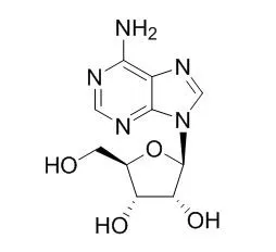
Adenosine is a nucleoside composed of a molecule of adenine attached to a ribose sugar molecule (ribofuranose) moiety via a β-N9-glycosidic bond. Adenosine induces SphK1 activity in human and mouse sickle and normal erythrocytes in vitro, it can activate the neuroimmune system, alter neuronal function and neurotransmission,and contribute to symptoms of sickness and psychopathologies. Adenosine activates mast cells have been long implicated in allergic asthma and studies in rodent mast cells have assigned the A3 Adenosine receptor (A3R) a primary role in mediating Adenosine responses.
|
| Targets: |
ERK | IL Receptor | VEGFR | cAMP | A3 Adenosine receptor | SphK1 |
| In vitro: |
| Mol Immunol. 2015 May;65(1):25-33. | | Down-regulation of the A3 adenosine receptor in human mast cells upregulates mediators of angiogenesis and remodeling.[Pubmed: 25597247] | Adenosine activated mast cells have been long implicated in allergic asthma and studies in rodent mast cells have assigned the A3 Adenosine receptor (A3R) a primary role in mediating Adenosine responses.
METHODS AND RESULTS:
Here we analyzed the functional impact of A3R activation on genes that are implicated in tissue remodeling in severe asthma in the human mast cell line HMC-1 that shares similarities with lung derived human mast cells. Quantitative real time PCR demonstrated upregulation of IL6, IL8, VEGF, amphiregulin and osteopontin. Moreover, further upregulation of these genes was noted upon the addition of dexamethasone. Unexpectedly, activated A3R down regulated its own expression and knockdown of the receptor replicated the pattern of agonist induced gene upregulation.
CONCLUSIONS:
This study therefore identifies the human mast cell A3R as regulator of tissue remodeling gene expression in human mast cells and demonstrates a heretofore-unrecognized mode of feedback regulation that is exerted by this receptor. | | Oncol Rep . 2019 Feb;41(2):829-838. | | Inhibition of autophagy enhances adenosine‑induced apoptosis in human hepatoblastoma HepG2 cells[Pubmed: 30535464] | | Abstract
In cancer research, autophagy acts as a double‑edged sword: it increases cell viability or induces cell apoptosis depending upon the cell context and functional status. Recent studies have shown that Adenosine (Ado) has cytotoxic effects in many tumors. However, the role of autophagy in Ado‑induced apoptosis is still poorly understood. In the present study, Ado‑induced apoptotic death and autophagy in hepatoblastoma HepG2 cells was investigated and the relationship between autophagy and apoptosis was identified. In the present study, it was demonstrated that Ado inhibited HepG2 cell growth in a time‑ and concentration‑dependent manner and activated endoplasmic reticulum (ER) stress, as indicated by G0/G1 cell cycle arrest, the increased mRNA and protein levels of GRP78/BiP, PERK, ATF4, CHOP, cleaved caspase‑3, cytochrome c and the loss of mitochon-drial membrane potential (ΔΨm). Ado also induced autophagic flux, revealed by the increased expression of the autophagy marker microtubule‑associated protein 1 light chain 3‑II (LC3‑II), Beclin‑1, autophagosomes, and the degradation of p62, as revealed by western blot analysis and macrophage‑derived chemokine (MDC) staining. Blocking autophagy using LY294002 notably entrenched Ado‑induced growth inhibition and cell apoptosis, as demonstrated with the increased expression of cytochrome c and p62, and the decreased expression of LC3‑II. Conversely, the autophagy inducer rapamycin alleviated Ado‑induced apoptosis and markedly increased the ΔΨm. Moreover, knockdown of AMPK with si‑AMPK partially abolished Ado‑induced ULK1 activation and mTOR inhibition, and thus reinforced CHOP expression and Ado‑induced apoptosis. These results indicated that Ado‑induced ER stress resulted in apoptosis and autophagy concurrently. The AMPK/mTOR/ULK1 signaling pathway played a protective role in the apoptotic procession. Inhibition of autophagy may effectively enhance the anticancer effect of Ado in human hepatoblastoma HepG2 cells. |
|
| In vivo: |
| J Neurochem. 2015 Jan;132(1):51-60. | | Adenosine transiently modulates stimulated dopamine release in the caudate-putamen via A1 receptors.[Pubmed: 25219576] | Adenosine modulates dopamine in the brain via A1 and A2A receptors, but that modulation has only been characterized on a slow time scale. Recent studies have characterized a rapid signaling mode of Adenosine that suggests a possible rapid modulatory role.
METHODS AND RESULTS:
Here, fast-scan cyclic voltammetry was used to characterize the extent to which transient Adenosine changes modulate stimulated dopamine release (5 pulses at 60 Hz) in rat caudate-putamen brain slices. Exogenous Adenosine was applied and dopamine concentration monitored. Adenosine only modulated dopamine when it was applied 2 or 5 s before stimulation. Longer time intervals and bath application of 5 μM Adenosine did not decrease dopamine release. Mechanical stimulation of endogenous Adenosine 2 s before dopamine stimulation also decreased stimulated dopamine release by 41 ± 7%, similar to the 54 ± 6% decrease in dopamine after exogenous Adenosine application. Dopamine inhibition by transient Adenosine was recovered within 10 min. The A1 receptor antagonist 8-cyclopentyl-1,3-dipropylxanthine blocked the dopamine modulation, whereas dopamine modulation was unaffected by the A2A receptor antagonist SCH 442416. Thus, transient Adenosine changes can transiently modulate phasic dopamine release via A1 receptors. These data demonstrate that Adenosine has a rapid, but transient, modulatory role in the brain. Here, transient Adenosine was shown to modulate phasic dopamine release on the order of seconds by acting at the A1 receptor. However, sustained increases in Adenosine did not regulate phasic dopamine release.
CONCLUSIONS:
This study demonstrates for the first time a transient, neuromodulatory function of rapid Adenosine to regulate rapid neurotransmitter release. | | Blood. 2015 Mar 5;125(10):1643-52. | | Elevated adenosine signaling via adenosine A2B receptor induces normal and sickle erythrocyte sphingosine kinase 1 activity.[Pubmed: 25587035] | Erythrocyte possesses high sphingosine kinase 1 (SphK1) activity and is the major cell type supplying plasma sphingosine-1-phosphate, a signaling lipid regulating multiple physiological and pathological functions.
Recent studies revealed that erythrocyte SphK1 activity is upregulated in sickle cell disease (SCD) and contributes to sickling and disease progression. However, how erythrocyte SphK1 activity is regulated remains unknown.
METHODS AND RESULTS:
Here we report that Adenosine induces SphK1 activity in human and mouse sickle and normal erythrocytes in vitro. Next, using 4 Adenosine receptor-deficient mice and pharmacological approaches, we determined that the A2B Adenosine receptor (ADORA2B) is essential for Adenosine-induced SphK1 activity in human and mouse normal and sickle erythrocytes in vitro. Subsequently, we provide in vivo genetic evidence that Adenosine deaminase (ADA) deficiency leads to excess plasma Adenosine and elevated erythrocyte SphK1 activity. Lowering Adenosine by ADA enzyme therapy or genetic deletion of ADORA2B significantly reduced excess Adenosine-induced erythrocyte SphK1 activity in ADA-deficient mice. Finally, we revealed that protein kinase A-mediated extracellular signal-regulated kinase 1/2 activation functioning downstream of ADORA2B underlies Adenosine-induced erythrocyte SphK1 activity.
CONCLUSIONS:
Overall, our findings reveal a novel signaling network regulating erythrocyte SphK1 and highlight innovative mechanisms regulating SphK1 activity in normal and SCD. | | Metabolism. 2014 Dec;63(12):1491-8. | | Modulation of neuroimmunity by adenosine and its receptors: metabolism to mental illness.[Pubmed: 25308443] | Adenosine is a pleiotropic bioactive with potent neuromodulatory properties. Due to its ability to easily cross the blood-brain barrier, it can act as a signaling molecule between the periphery and the brain. It functions through four (A1, A2A, A2B, and A3) cell surface G protein-coupled Adenosine receptors (ARs) that are expressed in some combination on nearly all cells types within the CNS. By regulating the activity of adenylyl cyclase and changing the intracellular concentration of cAMP, Adenosine can alter neuronal function and neurotransmission. A variety of illnesses related to metabolic dysregulation, such as type 1 diabetes and Alzheimer's disease, are associated with an elevated serum concentration of Adenosine and a pathogenesis rooted in inflammation.
CONCLUSIONS:
This review describes the accepted physiologic function of Adenosine in neurological disease and explores its new potential as a peripheral to central danger signal that can activate the neuroimmune system and contribute to symptoms of sickness and psychopathologies. |
|


 Cell. 2018 Jan 11;172(1-2):249-261.e12. doi: 10.1016/j.cell.2017.12.019.IF=36.216(2019)
 Cell Metab. 2020 Mar 3;31(3):534-548.e5. doi: 10.1016/j.cmet.2020.01.002.IF=22.415(2019)
 Mol Cell. 2017 Nov 16;68(4):673-685.e6. doi: 10.1016/j.molcel.2017.10.022.IF=14.548(2019)