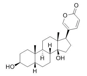
| In vitro: |
| J Tradit Chin Med. 2014 Dec;34(6):678-83. | | Reversal effect of bufalin on multidrug resistance in K562/VCR vincristine-resistant leukemia cell line.[Pubmed: 25618972] | To probe insights into the reversal effect of Bufalin on vincristine-acquired multidrug resistance (MDR) in human leukemia cell line K562/VCR.
METHODS AND RESULTS:
Proliferative inhibition rate and the reversal index (RI) of Bufalin were determined by Methyl thiazolyl tetrazolium assay. The uptake of Adriamycin (ADM) in K562/VCR cells, cell cycle and apoptosis rate were determined by flow cytometry (FCM). Cell morphologic changes were observed with Wright-Giemsa staining. The expression of P-glycoprotein (P-gp), multidrug-associated protein-1 (MRP1), Bcl-xL and Bax protein were measured by immunocytochemistry. The human leukemia multidrug resistant K562/VCR cells showed no cross-resistance to Bufalin. The RIs of Bufalin at concentrations of 0.0002, 0.001 and 0.005 μmol/L were 4.85, 6.94 and 14.77, respectively. Preincubation of 0.001 μmol/L Bufalin for 2 h could increase intracellular ADM fluorescence intensity to 28.07% (P < 0.05) and down-regulate MRP1 expression simultaneously, but no remarkable effect was found on P-gp protein. Cell cycle analysis indicated increased apoptosis rate and apparent decreased G2/M phase proportion after treatment with Bufalin. When exposed to 0.01 μmol/L Bufalin, typical morphological changes of apoptosis could be observed. Down-regulation of Bcl-xL and up-regulation of Bax expression in K562/VCR cells could be detected by immunocytochemistry.
CONCLUSIONS:
Bufalin could partly reverse the MDR of K562/VCR cells, with a possible mechanism of down-regulating MRP1 expression and activating apoptosis pathway by altering Bcl-xL/Bax ratio. | | J Biol Chem. 1996 Jun 14;271(24):14067-72. | | The cooperative interaction of two different signaling pathways in response to bufalin induces apoptosis in human leukemia U937 cells.[Pubmed: 8662906] | Bufalin, an active principle of Chinese medicine, chan'su, induced typical apoptosis in human leukemia U937 cells.
METHODS AND RESULTS:
When U937 cells were treated with 10(-8) M Bufalin in the absence of serum, mitogen-activated protein (MAP) kinase activity was markedly increased 6 h after the start of treatment and elevated so for 12 h. Prior to the activation of MAP kinase, increased activities of Ras, Raf-1, and MAP kinase kinase were found, but these enzymes were transiently activated by the treatment with Bufalin. These results suggest that the signal was transmitted sequentially from Ras, Raf-1, and MAP kinase kinase to MAP kinase. In association with this signal transduction, the concentration of cAMP in the cells decreased markedly, suggesting that Raf-1 was also activated by a decrease in the extent of phosphorylation by protein kinase A. In fact, pretreatment of U937 cells with forskolin and 3-isobutyl-1-methylxanthine, which are known to increase the concentration of cAMP in the cells, and subsequent treatment with Bufalin resulted in a decrease in both Raf-1 activity and DNA fragmentation. To confirm the participation of MAP kinase in the apoptotic process, antisense cDNA for MAP kinase kinase 1 was expressed in U937 cells. The transformants were significantly resistant to both DNA fragmentation and cell death in response to Bufalin.
CONCLUSIONS:
Our findings suggest that a pathway with the persistent activation of MAP kinase in U937 cells in response to Bufalin is at least one of the signal transduction pathways involved in the induction of apoptosis. |
|
| In vivo: |
| Environ Toxicol . 2017 Apr;32(4):1305-1317. | | Bufalin induces apoptosis in vitro and has Antitumor activity against human lung cancer xenografts in vivo[Pubmed: 27444971] | | Abstract
Bufalin has been shown to be effective against a variety of cancer cells, but its role in lung cancer has never been studied in an animal model. In this study, we evaluated Bufalin effects in a human lung cancer cell line NCI-H460 both in vitro and in vivo. Bufalin caused significant cytotoxicity in NCI-H460 cells at a concentration as low as 1 μM. DNA condensation was observed in Bufalin-treated cells in a dose-dependent manner. Mitochondrial membrane potential (ΔΨm ) was reduced and reactive oxygen species (ROS) were increased in Bufalin-treated NCI-H460 cells. Levels of several proapoptotic proteins such as Fas, Fas-ligand, cytochrome c, apoptosis protease activating factor-1, endonuclease G, caspase-3 and caspase-9 were increased after Bufalin treatment. At the same time, anti-apoptotic B-cell lymphoma 2 protein levels were reduced. Bufalin decreased glucose regulated protein-78 gene expression but increased growth arrest- and DNA damage-inducible 153 gene expression. Bufalin injected intraperitoneally in a dose-dependent manner reduced tumor size in BALB/C nu/nu mice implanted with NCI-H460 cells. Bufalin injection did not produce significant drug-related toxicity in experimental animals except at a high dose (0.4 mg kg-1 ). In conclusion, low concentrations of Bufalin can induce apoptosis in the human lung cancer cell line NCI-H460 in vitro. Bufalin also reduced tumor size in mice injected with NCI-H460 cells without significant drug-related toxicity. These results indicate that Bufalin may have potential to be developed as an agent for treating human non-small cell lung cancer. © 2016 Wiley Periodicals, Inc. Environ Toxicol 32: 1305-1317, 2017.
Keywords: animal models; apoptosis; Bufalin; lung neoplasm. |
|


 Cell. 2018 Jan 11;172(1-2):249-261.e12. doi: 10.1016/j.cell.2017.12.019.IF=36.216(2019)
 Cell Metab. 2020 Mar 3;31(3):534-548.e5. doi: 10.1016/j.cmet.2020.01.002.IF=22.415(2019)
 Mol Cell. 2017 Nov 16;68(4):673-685.e6. doi: 10.1016/j.molcel.2017.10.022.IF=14.548(2019)