| In vitro: |
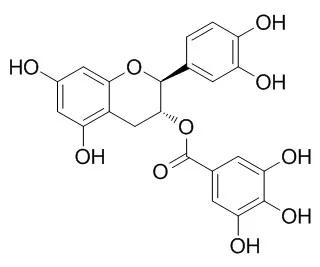
| Toxicol Lett. 2007 Jul 10;171(3):171-80. | | In vitro cytotoxicity of (-)-catechin gallate, a minor polyphenol in green tea.[Pubmed: 17606338] | The cytotoxicity of (-)-Catechin gallate(CG), a minor polyphenolic constituent in green tea, towards cells derived from tissues of the human oral cavity was studied.
METHODS AND RESULTS:
The sequence of sensitivity to (-)-Catechin gallate(CG) was: immortalized epithelioid gingival S-G cells>tongue squamous carcinoma CAL27 cells>salivary gland squamous carcinoma HSG cells>>normal gingival HGF-1 fibroblasts. Further studies focused on S-G cells, the cells most sensitive to (-)-Catechin gallate(CG). The response of the S-G cells to (-)-Catechin gallate(CG) was dependent on the length of exposure, with midpoint cytotoxicity values of 127, 67 and 58muM (-)-Catechin gallate(CG) for 1-, 2- and 3-day exposures, respectively. The sequence of sensitivity of the S-G cells to various green tea catechins was characterized as follows: (-)-Catechin gallate(CG), epicatechin gallate (ECG)>epigallocatechin gallate (EGCG)>epigallocatechin (EGC)>>epicatechin (EC), catechin (C). The cytotoxicity of (-)-Catechin gallate(CG), apparently, was not due to oxidative stress as it was a poor generator of H(2)O(2) in tissue culture medium, had no effect on the intracellular glutathione level, its cytotoxicity was unaffected by catalase, and it did not induce lipid peroxidation. However, (-)-Catechin gallate(CG) did enhance Fe(2+)-induced, lipid peroxidation. (-)-Catechin gallate(CG)-induced apoptosis was detected by nuclear staining, both with acridine orange and by the more specific TUNEL procedure. The lack of caspase-3 activity in cells exposed to (-)-Catechin gallate(CG) and the detection of a DNA smear, rather than of discrete internucleosomal DNA fragmentation, upon agarose gel electrophoresis, suggest, possibly, that the mode of cell death was by a caspase-independent apoptotic pathway.
CONCLUSIONS:
The overall cytotoxicity of (-)-Catechin gallate(CG) was similar to its epimer, ECG and both exhibited antiproliferative effects equivalent to, or stronger than, EGCG, the most abundant catechin in green tea. | | Food Chem.,2010,122(4):1061-6. | | In vitro inhibition of α-glucosidases and glycogen phosphorylase by catechin gallates in green tea.[Reference: WebLink] | We investigated in vitro inhibition of mammalian carbohydrate-degrading enzymes by green tea extract and the component catechins, and further evaluated their inhibitory activities in cell cultures.
METHODS AND RESULTS:
The extract showed good inhibition toward rat intestinal maltase and rabbit glycogen phosphorylase (GP) b, with IC50 values of 45 and 7.4 μg/ml, respectively. The polyphenol components, catechin 3-gallate (CG), gallocatechin 3-gallate (GCG), epicatechin 3-gallate (ECG), and epigallocatechin 3-gallate (EGCG), were good inhibitors of maltase, with IC50 values of 62, 67, 40, and 16 μM, respectively, and EGCG also showed good inhibition toward maltase expressed on Caco-2 cells, with an IC50 value of 27 μM. The ungallated catechins, such as catechin, gallocatechin (GC), epicatechin (EC), and epigallocatechin (EGC), showed no significant inhibition toward GP b, whereas the gallated catechins CG, GCG, ECG, and EGCG inhibited the enzyme, with IC50 values of 35, 6.3, 27, and 34 μM. From multiple inhibition studies by Dixon plots, GCG appears to bind a new allostelic site, the indole inhibitor site. These gallated catechins also inhibited glucagon-stimulated glucose production dose-dependently, with IC50 values ranging from 33 to 55 μM.
CONCLUSIONS:
Dietary supplementation with these gallated catechins or the green tea extract containing them, which inhibits both α-glucosidases and GP in vitro and in cell culture, would contribute to the protection or improvement of type 2 diabetes. |
|


 Cell. 2018 Jan 11;172(1-2):249-261.e12. doi: 10.1016/j.cell.2017.12.019.IF=36.216(2019)
 Cell Metab. 2020 Mar 3;31(3):534-548.e5. doi: 10.1016/j.cmet.2020.01.002.IF=22.415(2019)
 Mol Cell. 2017 Nov 16;68(4):673-685.e6. doi: 10.1016/j.molcel.2017.10.022.IF=14.548(2019)