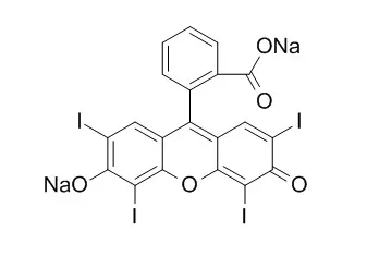
| In vitro: |
| Biotechniques . 2020 Jan;68(1):7-13. | | Erythrosin B: a versatile colorimetric and fluorescent vital dye for bacteria[Pubmed: 31718252] | | Rapidly assaying cell viability for diverse bacteria species is not always straightforward. In eukaryotes, cell viability is often determined using colorimetric dyes; however, such dyes have not been identified for bacteria. We screened different dyes and found that Erythrosin B (EB), a visibly red dye with fluorescent properties, functions as a vital dye for many Gram-positive and -negative bacteria. EB worked at a similar concentration for all bacteria studied and incubations were as short as 5 min. Given EB's spectral properties, diverse experimental approaches are possible to rapidly visualize and/or quantitate dead bacterial cells in a population. As the first broadly applicable colorimetric viability dye for bacteria, EB provides a cost-effective alternative for researchers in academia and industry. | | Antiviral Res . 2018 Feb;150:217-225. | | Erythrosin B is a potent and broad-spectrum orthosteric inhibitor of the flavivirus NS2B-NS3 protease[Pubmed: 29288700] | | Many flaviviruses, such as Zika virus (ZIKV), Dengue virus (DENV1-4) and yellow fever virus (YFV), are significant human pathogens. Infection with ZIKV, an emerging mosquito-borne flavivirus, is associated with increased risk of microcephaly in newborns and Guillain-Barré syndrome and other complications in adults. Currently, specific therapy does not exist for any flavivirus infections. In this study, we found that Erythrosin B, an FDA-approved food additive, is a potent inhibitor for flaviviruses, including ZIKV and DENV2. Erythrosin B was found to inhibit the DENV2 and ZIKV NS2B-NS3 proteases with IC50 in low micromolar range, via a non-competitive mechanism. Erythrosin B can significantly reduce titers of representative flaviviruses, DENV2, ZIKV, YFV, JEV, and WNV, with micromolar potency and with excellent cytotoxicity profile. Erythrosin B can also inhibit ZIKV replication in ZIKV-relevant human placental and neural progenitor cells. As a pregnancy category B food additive, Erythrosin B may represent a promising and easily developed therapy for management of infections by ZIKV and other flaviviruses. | | J Histochem Cytochem . 1984 Oct;32(10):1084-1090. | | Fluorescent erythrosin B is preferable to trypan blue as a vital exclusion dye for mammalian cells in monolayer culture[Pubmed: 6090533] | | Erythrosin B and trypan blue are tested and compared for their effectiveness as vital exclusion stains for mammalian cells in monolayer culture. Both stains are supposed to mark cells that have lost membrane integrity. Fluorescein diacetate (FDA), an efficient vital inclusion stain, is used as a control, as it marks cells retaining membrane integrity. Erythrosin B and FDA are used as fluorescent dyes, whereas trypan blue colors via light absorption. The effectiveness of both vital exclusion stains is assayed by their ability to stain a high percentage of monolayer cells exposed to treatments lethal to an entire cell population. Two types of lethal treatment, severe heat and metabolic poison, are employed. Erythrosin B stains all monolayer cells immediately after complete lethal treatment. Trypan blue optimally stains only about 60% of monolayer cells. Cell staining by Erythrosin B and by FDA are found to be mutually exclusive. This result demonstrates the coincidence of viability indications by Erythrosin B and FDA and thus confirms the reliability of both viability stains as they probe membrane permeability via independent mechanisms. This study shows that Erythrosin B is an effective, nontoxic, and convenient fluorescent vital exclusion dye for three mammalian cell lines in monolayer culture, but tends to disqualify trypan blue for this application. | | Food Res Int . 2017 Oct;100(Pt 1):344-351. | | Enhanced antimicrobial effect of ultrasound by the food colorant Erythrosin B[Pubmed: 28873696] | | The synergistic combination of the food colorant Erythrosin B (E-B, FD&C 3) (0, 25, and 50μM) and low-frequency ultrasound (20kHz, 0.86-0.90WmL-1) was evaluated against Listeria innocua. Although E-B was antibacterial by itself, the inactivation rate significantly increased in a concentration-dependent manner upon exposure to ultrasound and followed a sigmoidal behavior. The enhanced antimicrobial effect of E-B in the presence of ultrasound can be explained in part from a microbubble disappearance study in which it was confirmed that the presence of E-B enhances inertial cavitation, thereby enhancing the antimicrobial effect of ultrasound. The inactivation rate in a sequential treatment, where L. innocua was sonicated for 4min followed by exposure to 25μM Erythrosin B, was comparable to that obtained by the simultaneous treatment, indicating complementary mechanisms of inactivation. Fluorescence microscopy showed attachment of E-B to the cells, which may explain its intrinsic antimicrobial property. Other mechanism may include the confirmed decrease in the cavitation threshold of water by addition of E-B, resulting in more effective cavitation. The study offers a proof-of-concept of a novel approach to complement ultrasound treatment for enhanced microbial inactivation. | | J Physiol . 1983 Jan;334:47-63. | | Neurotransmitter release and nerve terminal morphology at the frog neuromuscular junction affected by the dye Erythrosin B[Pubmed: 6134825] | | 1. The quantal release of neurotransmitter and the fine structure of frog neuromuscular junctions has been examined in the presence of the xanthene dye Erythrosin B.2. At concentrations of 10 muM or greater, Erythrosin B produced time- and dose-dependent increases in transmitter release from presynaptic nerve terminals.3. Miniature end-plate potential (m.e.p.p.) frequency increased in an exponential manner during continuous exposure to the dye. The rate constant for this exponential was dose-dependent, increasing with concentrations from 10 muM to 1 mM.4. The amplitude of evoked end-plate potentials (e.p.p.s) also increased exponentially during dye treatment, primarily due to an increase in quantal content. Rate constants for this effect were also dose-dependent, and were approximately 1/5 as large as those for m.e.p.p.s.5. While the frequency of m.e.p.p.s was increasing, their amplitude distribution did not qualitatively change. Thus the dye has little effect on the size of individual quanta.6. The presynaptic effects of Erythrosin B were irreversible under these experimental conditions. Brief exposure to the dye caused increases in m.e.p.p. frequency and e.p.p. amplitude which were maintained at steady levels during extensive rinsing with dye-free Ringer solution.7. Prolonged exposure to the dye caused an eventual decrease in m.e.p.p. frequency and abolition of e.p.p.s. Coincident with this decline ;giant' m.e.p.p.s as large as 40 mV were observed.8. At dye concentrations greater than approximately 200 muM, Erythrosin B rapidly and reversibly increased the membrane potential and input resistance of muscle fibres. This post-synaptic effect was small and variable in normal saline, but was pronounced in low potassium solutions.9. During the period that release was enhanced by Erythrosin B, presynaptic nerve terminals contained the normal complement of synaptic vesicles and other organelles. Mitochondria were swollen in this condition.10. After m.e.p.p. frequency declined below normal levels and ;giant' m.e.p.p.s appeared, the number of synaptic vesicles within nerve terminals declined and dilated cisternae were present. Mitochondria were swollen further.11. These results do not reveal any mechanism to explain the ability of Erythrosin B to increase transmitter release, but the decline in release may be caused by partial depletion of synaptic vesicles. The ;giant' m.e.p.p.s could be due to the discharge of acetylcholine from cisternae. |
|


 Cell. 2018 Jan 11;172(1-2):249-261.e12. doi: 10.1016/j.cell.2017.12.019.IF=36.216(2019)
 Cell Metab. 2020 Mar 3;31(3):534-548.e5. doi: 10.1016/j.cmet.2020.01.002.IF=22.415(2019)
 Mol Cell. 2017 Nov 16;68(4):673-685.e6. doi: 10.1016/j.molcel.2017.10.022.IF=14.548(2019)