| In vitro: |
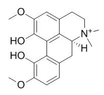
| J Nat Med. 2015 Apr 4. | | Sinomenine and magnoflorine, major constituents of Sinomeni Caulis et Rhizoma, show potent protective effects against membrane damage induced by lysophosphatidylcholine in rat erythrocytes.[Pubmed: 25840917 ] | The effects of the water extract of Sinomeni Caulis et Rhizoma (SCR-WE) and its major constituents, sinomenine (SIN) and Magnoflorine (MAG), on moderate hemolysis induced by lysophosphatidylcholine (LPC) were investigated in rat erythrocytes and compared with the anti-hemolytic effects of lidocaine (LID) and propranolol (PRO) as reference drugs.
METHODS AND RESULTS:
LPC caused hemolysis at concentrations above the critical micelle concentration (CMC), and the concentration of LPC producing moderate hemolysis (60 %) was approximately 10 μM. SCR-WE at 1 ng/mL-100 μg/mL significantly inhibited the hemolysis induced by LPC. SIN and Magnoflorine attenuated LPC-induced hemolysis in a concentration-dependent manner from very low to high concentrations (1 nM-100 μM and 10 nM-100 μM, respectively). In contrast, the inhibiting effects of LID and PRO on LPC-induced hemolysis were observed at higher concentrations (1-100 μM) but not at lower concentrations (1-100 nM). Neither SIN nor Magnoflorine affected micelle formation of LPC, nor, at concentrations of 1 nM-1 μM, did they attenuate the hemolysis induced by osmotic imbalance (hypotonic hemolysis). Similarly, SCR-WE also did not modify micelle formation or hypotonic hemolysis, except at the highest concentration.
CONCLUSIONS:
These results suggest that SIN and Magnoflorine potently protect the erythrocyte membrane from LPC-induced damage and contribute to the beneficial action of SCR-WE. The protective effects of SIN and Magnoflorine are mediated by some mechanism other than prevention of micelle formation or protection of the erythrocyte membrane against osmotic imbalance. | | Biol Pharm Bull. 2007 Jun;30(6):1157-60. | | Magnoflorine from Coptidis Rhizoma protects high density lipoprotein during oxidant stress.[Pubmed: 17541173] | The objective of the present study was to investigate the beneficial properties of Magnoflorine, an alkaloid isolated from coptidis rhizoma, on protecting human high density lipoprotein (HDL) against lipid peroxidation.
METHODS AND RESULTS:
Magnoflorine exerts an inhibitory effect against Cu2+-induced lipid peroxidation of HDL, as showed by prolongation of lag time from 62 to 123 min at the concentration of 3.0 microM. It also inhibits the generation of thiobarbituric acid reactive substances (TBARS) in the dose-dependent manners with IC50 values of 2.3+/-0.2 microM and 6.2+/-0.5 microM since HDL oxidation mediated by either catalytic Cu2+ or thermo-labile radical initiator (AAPH), respectively. Separately, Cu2+ oxidized HDL lost the antioxidant action but the inclusion of Magnoflorine/Cu2+ oxidized HDL can protect LDL oxidation according to increasing Magnoflorine concentration.
CONCLUSIONS:
The results suggest that Magnoflorine may have a role to play in preventing the HDL oxidation. | | World J Microbiol Biotechnol . 2018 Oct 31;34(11):167. | | Antifungal activity of magnoflorine against Candida strains[Pubmed: 30382403] | | Abstract
Candida albicans is a major invasive pathogen, and the development of strains resistant to conventional antifungal agents has been reported in recent years. We evaluated the antifungal activity of 44 compounds against Candida strains. Magnoflorine showed the highest growth inhibitory activity of the tested Candida strains, with a minimum inhibitory concentration (MIC) of 50 μg/mL based on microdilution antifungal susceptibility testing. Disk diffusion assay confirmed the antifungal activity of Magnoflorine and revealed that this activity was stable over 3 days compared to those of berberine and cinnamaldehyde. Cytotoxicity testing showed that Magnoflorine could potentially be used in a clinical setting because it didn't have any toxicity to HaCaT cells even in 200 μg/mL of treatment. Magnoflorine at 50 μg/mL inhibited 55.91 ± 7.17% of alpha-glucosidase activity which is required for normal cell wall composition and virulence of Candida albicans. Magnoflorine also reduced the formation of C. albicans' biofilm. Combined treatment with Magnoflorine and miconazole decreased the amount of miconazole required to kill various Candida albicans. Therefore, Magnoflorine is a good candidate lead compound for novel antifungal agents.
Keywords: Alpha-glucosidase; Antifungal; Candida albicans; Magnoflorine; Susceptibility microdilution assay. | | Planta Med . 2018 Nov;84(17):1255-1264. | | Magnoflorine Enhances LPS-Activated Pro-Inflammatory Responses via MyD88-Dependent Pathways in U937 Macrophages[Pubmed: 29906814] | | Abstract
Magnoflorine, a major bioactive metabolite isolated from Tinospora crispa, has been reported for its diverse biochemical and pharmacological properties. However, there is little report on its underlying mechanisms of action on immune responses, particularly on macrophage activation. In this study, we aimed to investigate the effects of Magnoflorine, isolated from T. crispa on the pro-inflammatory mediators generation induced by LPS and the concomitant NF-κB, MAPKs, and PI3K-Akt signaling pathways in U937 macrophages. Differentiated U937 macrophages were treated with Magnoflorine and the release of pro-inflammatory mediators was evaluated through ELISA, while the relative mRNA expression of the respective mediators was quantified through qRT-PCR. Correspondingly, western blotting was executed to observe the modulatory effects of Magnoflorine on the expression of various markers related to NF-κB, MAPK and PI3K-Akt signaling activation in LPS-primed U937 macrophages. Magnoflorine significantly enhanced the upregulation of TNF-α, IL-1β, and PGE2 production as well as COX-2 protein expression. Successively, Magnoflorine prompted the mRNA transcription level of these pro-inflammatory mediators. Magnoflorine enhanced the NF-κB activation by prompting p65, IκBα, and IKKα/β phosphorylation as well as IκBα degradation. Besides, Magnoflorine treatments concentration-dependently augmented the phosphorylation of JNK, ERK, and p38 MAPKs as well as Akt. The immunoaugmenting effects were further confirmed by investigating the effects of Magnoflorine on specific inhibitors, where the treatment with specific inhibitors of NF-κB, MAPKs, and PI3K-Akt proficiently blocked the Magnoflorine-triggered TNF-α release and COX-2 expression. Magnoflorine furthermore enhanced the MyD88 and TLR4 upregulation. The results suggest that Magnoflorine has high potential on augmenting immune responses. |
|


 Cell. 2018 Jan 11;172(1-2):249-261.e12. doi: 10.1016/j.cell.2017.12.019.IF=36.216(2019)
 Cell Metab. 2020 Mar 3;31(3):534-548.e5. doi: 10.1016/j.cmet.2020.01.002.IF=22.415(2019)
 Mol Cell. 2017 Nov 16;68(4):673-685.e6. doi: 10.1016/j.molcel.2017.10.022.IF=14.548(2019)