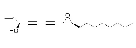
| Kinase Assay: |
| Apoptosis. 2011 Apr;16(4):347-58. | | Panaxydol induces apoptosis through an increased intracellular calcium level, activation of JNK and p38 MAPK and NADPH oxidase-dependent generation of reactive oxygen species.[Pubmed: 21190085 ] | Panaxydol, a polyacetylenic compound derived from Panax ginseng roots, has been shown to inhibit the growth of cancer cells.
METHODS AND RESULTS:
In this study, we demonstrated that Panaxydol induced apoptosis preferentially in transformed cells with a minimal effect on non-transformed cells. Furthermore, Panaxydol was shown to induce apoptosis through an increase in intracellular Ca(2+) concentration ([Ca(2+)](i)), activation of JNK and p38 MAPK, and generation of reactive oxygen species (ROS) initially by NADPH oxidase and then by mitochondria. Panaxydol-induced apoptosis was caspase-dependent and occurred through a mitochondrial pathway. ROS generation by NADPH oxidase was critical for Panaxydol-induced apoptosis. Mitochondrial ROS production was also required, however, it appeared to be secondary to the ROS generation by NADPH oxidase. Activation of NADPH oxidase was demonstrated by the membrane translocation of regulatory p47(phox) and p67(phox) subunits and shown to be necessary for ROS generation by Panaxydol treatment. Panaxydol triggered a rapid and sustained increase of [Ca(2+)](i), which resulted in activation of JNK and p38 MAPK. JNK and p38 MAPK play a key role in activation of NADPH oxidase, since inhibition of their expression or activity abrogated membrane translocation of p47(phox) and p67(phox) subunits and ROS generation.
CONCLUSIONS:
In summary, these data indicate that Panaxydol induces apoptosis preferentially in cancer cells, and the signaling mechanisms involve a [Ca(2+)](i) increase, JNK and p38 MAPK activation, and ROS generation through NADPH oxidase and mitochondria. | | J Clin Neurosci. 2009 Mar;16(3):444-8. | | Phosphatidylinositol 3-kinase activity is required for the induction of differentiation in C6 glioma cells by panaxydol.[Pubmed: 19179079] | Panaxydol isolated from the lipophilic fractions of Tienchi ginseng (Panax notoginseng) induces growth inhibition and differentiation of rat C6 glioma cells. The underlying molecular mechanisms are not completely understood.
METHODS AND RESULTS:
In the present study, we identified phosphatidylinositol 3-kinase (PI 3-K) as a necessary enzyme for the differentiation of C6 cells treated with Panaxydol. The specific PI 3-K inhibitor wortmannin resulted in attenuated differentiation of C6 cells induced by Panaxydol, and was associated with perinuclear localization of glial fibrillary acidic protein expression and a diminished process formation.
CONCLUSIONS:
These data suggest that induction of differentiation in C6 cells by Panaxydol could be mediated through a PI 3-K-dependent pathway. |
|
| Cell Research: |
| Chem Biol Interact. 2009 Sep 14;181(1):138-43. | | Panaxydol inhibits the proliferation and induces the differentiation of human hepatocarcinoma cell line HepG2.[Pubmed: 19450571] | Panaxydol, a polyacetylene compound isolated from Panax ginseng, exerts anti-proliferative effects against malignant cells. No previous study, however, has been reported on its effects on hepatocellular carcinoma cells.
METHODS AND RESULTS:
Here, we investigated the effects of Panaxydol on the proliferation and differentiation of human hepatocarcinoma cell line HepG2. We studied by electronic microscopy of morphological and ultrastructural changes induced by Panaxydol. We also examined the cytotoxicities of Panaxydol against HepG2 cells using the 3-(4,5-dimethyl-2-thiazolyl)-2,5-diphenyl tetrazolium bromide assay and the effect of Panaxydol on cell cycle distributions by flow cytometry. We investigated the production of liver proteins in Panaxydol-treated cells including alpha-fetoprotein and albumin and measured the specific activity of alkaline phosphatase and gamma-glutamyl transferase. We further investigated the effects of Panaxydol on the expression of Id-1, Id-2, p21 and pRb by RT-PCR or immunoblotting analysis. We found that Panaxydol inhibited the proliferation of HepG2 cells and caused morphological and ultrastructural changes in HepG2 cells resembling more mature forms of hepatocytes. Moreover, Panaxydol induced a cell cycle arrest at the G(1) to S transition in HepG2 cells. It also significantly decreased the secretion of alpha-fetoprotein and the activity of gamma-glutamyl transferase. By contrast, Panaxydol remarkably increased the secretion of albumin and the alkaline phosphatase activity. Furthermore, Panaxydol increased the mRNA content of p21 while reducing that of Id-1 and Id-2. Panaxydol also increased the protein levels of p21, pRb and the hypophosphorylated pRb in a dose-dependent manner.
CONCLUSIONS:
These findings suggest that Panaxydol is of value for further exploration as a potential anti-cancer agent. |
|


 Cell. 2018 Jan 11;172(1-2):249-261.e12. doi: 10.1016/j.cell.2017.12.019.IF=36.216(2019)
 Cell Metab. 2020 Mar 3;31(3):534-548.e5. doi: 10.1016/j.cmet.2020.01.002.IF=22.415(2019)
 Mol Cell. 2017 Nov 16;68(4):673-685.e6. doi: 10.1016/j.molcel.2017.10.022.IF=14.548(2019)