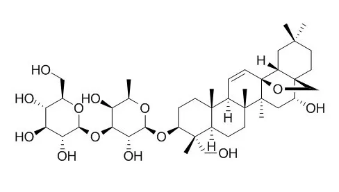
| Description: |
Saikosaponin D, a calcium mobilizing agent (SERCA inhibitor), is also an agonist of the glucocorticoid receptor (GR),which has anti-cancer, anti-inflammatory, and neuroprotective effects. Saikosaponin D protects against acetaminophen-induced hepatotoxicity by inhibiting NF-κB and STAT3 signaling; it shows inhibitory effects on NF-κB activation and thereby on iNOS, COX-2 and pro-inflammatory cytokines. |
| In vitro: |
| Eur Rev Med Pharmacol Sci. 2014;18(17):2435-43. | | Saikosaponin-d inhibits proliferation of human undifferentiated thyroid carcinoma cells through induction of apoptosis and cell cycle arrest.[Pubmed: 25268087] | Saikosaponin D is a triterpene saponin derived from Bupleurum falcatum. L and has been reported to exhibit a variety of pharmacological activities such as anti-bacterial, anti-virus and anti-cancer. The aim of the present study was to explore the effect of Saikosaponin D on the proliferation and apoptosis of human undifferentiated thyroid carcinoma.
METHODS AND RESULTS:
Three human anaplastic thyroid cancers cell lines were cultured in the presence of Saikosaponin D and their proliferation was measured by MTT assay. Cell apoptosis and cell cycle distribution were analyzed with flow cytometry. Western blot was performed to determine the proteins expression. The in vivo effect of Saikosaponin D was measured with an animal model.
In vitro, MTT assay showed that Saikosaponin D treatment inhibited cell proliferation in three human anaplastic thyroid cancers cell lines ARO, 8305C and SW1736. In addition, Saikosaponin D promoted cell apoptosis and induced G1-phase cell cycle arrest as shown by flow cytometric analysis. On the molecular level, our results showed that saikosaponin-d treatment increased the expression of p53 and bax, and decreased the expression of Bcl-2. In addition, saikosaponin-d administration led to a significant up-regulation of p21 and down-regulation of CDK2 and cyclin D1. Xenografts tumorigenesis model demonstrated that Saikosaponin D markedly reduced the weight and volume of thyroid tumors in vivo.
CONCLUSIONS:
The present study suggested that Saikosaponin D might be a new potent chemopreventive drug candidate for human undifferentiated thyroid carcinoma through induction of apoptosis and cell cycle arrest. | | Int. Immunopharmacol., 2012 Sep;14(1):121-6. | | Saikosaponin a and its epimer saikosaponin d exhibit anti-inflammatory activity by suppressing activation of NF-κB signaling pathway.[Pubmed: 22728095 ] | Saikosaponin a (SSa) and its epimer Saikosaponin D (SSd) are major triterpenoid saponin derivatives from Radix bupleuri (RB), which has been long used in Chinese traditional medicine for treatment of various inflammation-related diseases.
METHODS AND RESULTS:
In the present study, the anti-inflammatory activity, as well as the underlying mechanism, of SSa and SSd was investigated in lipopolysaccharide (LPS)-induced RAW264.7 cells. Our results demonstrated that both SSa and SSd significantly inhibited the expression of inducible nitric-oxide synthase (iNOS) and cyclooxygenase-2 (COX-2) in LPS-induced RAW264.7 cells, and finally resulted in the reduction of nitric oxide (NO) and prostaglandin E(2) (PGE(2)). In addition, LPS-induced production of major pro-inflammatory cytokines: the tumor necrosis factor-α (TNF-α) and interleukin-6 (IL-6), was suppressed in a dose-dependent manner by the treatment of SSa or SSd in RAW264.7 cells. Further analysis revealed that both SSa and SSd could inhibit translocation of nuclear factor-κB (NF-κB) from the cytoplasm to the nucleus in the LPS-induced RAW264.7 cells. Furthermore, SSa and SSd exhibited significant anti-inflammatory activity in two different murine models of acute inflammation, carrageenan-induced paw edema in rats and acetic acid-induced vascular permeability in mice.
CONCLUSIONS:
In conclusion, SSa and SSd showed potent anti-inflammatory activity through inhibitory effects on NF-κB activation and thereby on iNOS, COX-2 and pro-inflammatory cytokines. | | 2018 Mar;41(3):1357-1364. | | Estrogen receptor‑β‑dependent effects of saikosaponin‑d on the suppression of oxidative stress‑induced rat hepatic stellate cell activation[Pubmed: 29286085] | | Abstract
Saikosaponin-d (SSd) is one of the major triterpenoid saponins derived from Bupleurum falcatum L., which has been reported to possess antifibrotic activity. At present, there is little information regarding the potential target of SSd in hepatic stellate cells (HSCs), which serve an important role in excessive extracellular matrix (ECM) deposition during the pathogenesis of hepatic fibrosis. Our recent study indicated that SSd may be considered a novel type of phytoestrogen with estrogen‑like actions. Therefore, the present study aimed to investigate the effects of SSd on the proliferation and activation of HSCs, and the underlying mechanisms associated with estrogen receptors. In the present study, a rat HSC line (HSC‑T6) was used and cultured with dimethyl sulfoxide, SSd, or estradiol (E2; positive control), in the presence or absence of three estrogen receptor (ER) antagonists [ICI‑182780, methylpiperidinopyrazole (MPP) or (R,R)-tetrahydrochrysene (THC)], for 24 h as pretreatment. Oxidative stress was induced by exposure to hydrogen peroxide for 4 h. Cell proliferation was assessed by MTT growth assay. Malondialdehyde (MDA), CuZn-superoxide dismutase (CuZn-SOD), tissue inhibitor of metalloproteinases-1 (TIMP-1), matrix metalloproteinase-1 (MMP-1), transforming growth factor-β1 (TGF-β1), hydroxyproline (Hyp) and collagen-1 (COL1) levels in cell culture supernatants were determined by ELISA. Reactive oxygen species (ROS) was detected by flow cytometry. Total and phosphorylated mitogen-activated protein kinases (MAPKs) and α-smooth muscle actin (α-SMA) were examined by western blot analysis. TGF-β1 mRNA expression was determined by RT-quantitative (q)PCR. SSd and E2 were able to significantly suppress oxidative stress‑induced proliferation and activation of HSC‑T6 cells. Furthermore, SSd and E2 were able to reduce ECM deposition, as demonstrated by the decrease in transforming growth factor‑β1, hydroxyproline, collagen‑1 and tissue inhibitor of metalloproteinases‑1, and by the increase in matrix metalloproteinase‑1. These results suggested that the possible molecular mechanism could involve downregulation of the reactive oxygen species/mitogen‑activated protein kinases signaling pathway. Finally, the effects of SSd and E2 could be blocked by co‑incubation with ICI‑182780 or THC, but not MPP, thus indicating that ERβ may be the potential target of SSd in HSC‑T6 cells. In conclusion, these findings suggested that SSd may suppress oxidative stress‑induced activation of HSCs, which relied on modulation of ERβ. |
|


 Cell. 2018 Jan 11;172(1-2):249-261.e12. doi: 10.1016/j.cell.2017.12.019.IF=36.216(2019)
 Cell Metab. 2020 Mar 3;31(3):534-548.e5. doi: 10.1016/j.cmet.2020.01.002.IF=22.415(2019)
 Mol Cell. 2017 Nov 16;68(4):673-685.e6. doi: 10.1016/j.molcel.2017.10.022.IF=14.548(2019)