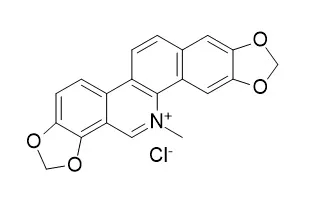
| In vitro: |
| Biochemistry & Biophysics Reports, 2018, 14:149–154. | | Natural compound sanguinarine chloride targets the type III secretion system of Salmonella enterica Serovar Typhimurium[Reference: WebLink] | The type III secretion system (T3SS) is a key virulence mechanism of many Gram-negative bacterial pathogens. Upon contact between bacteria and host cells, T3SS transfers a series of effectors from the bacterial cytosol to host cells. It is widely known that a mutation in T3SS does not impair bacterial growth, thereby avoiding any subsequent development of resistance. Thus, T3SS is expected to be a candidate therapeutic target.
METHODS AND RESULTS:
While developing the T3SS screening method, we discovered that Sanguinarine chloride, a natural compound, could decrease the production of the SPI-1 type III secretion system main virulence proteins SipA and SipB and prevent the invasion of HeLa cells by Salmonella enterica serovar Typhimurium without affecting the growth of Salmonella. Furthermore, Sanguinarine chloride downregulated the transcription of HilA and consequently regulated the expression of the SPI-1 apparatus and effector genes.
CONCLUSIONS:
In summary, our study directly demonstrated that this putative SPI-1 inhibitor belongs to a novel class of anti-Salmonella compounds. | | Pharmacol Toxicol. 1996 Jun;78(6):397-403. | | Cytotoxicity of sanguinarine chloride to cultured human cells from oral tissue.[Pubmed: 8829200] |
METHODS AND RESULTS:
The in vitro cytotoxicity of Sanguinarine chloride, a dental product used in the treatment of gingivitis and plaque, was compared using cell lines and primary cells from oral human tissues. For the established cell lines, Sanguinarine chloride exhibited similar potencies to S-G gingival epithelial cells and to KB carcinoma cells, whereas HGF-1 gingival fibroblasts were more tolerant. However, a gingival primary cell culture was more sensitive to Sanguinarine chloride than were the established cell lines. Detailed studies were performed with the S-G cells. The 24-hr midpoint (NR50) cytotoxicity value towards the S-G cells was 7.6 microM, based on the neutral red cytotoxicity assay; vacuolization and multinucleation were noted. When exposed to Sanguinarine chloride for 3 days, a lag in growth kinetics was first observed at 1.7 microM. Damage to the integrity of the plasma membrane was evident, as leakage of lactic acid dehydrogenase occurred during a 3 hr exposure to Sanguinarine chloride at 0.1275 mM and greater.
CONCLUSIONS:
The cytotoxicity of Sanguinarine chloride to the S-G cells was lessened in the presence of an S9 hepatic microsomal fraction from Aroclor-induced rats or by including fetal bovine serum (15%) in the exposure medium. Progressively increasing the pH from 6.0 to 7.8 enhanced the potency of Sanguinarine chloride, presumably due to the enhanced uptake of the lipophilic alkanolamine form, as compared to that of the cationic iminium form. |
|


 Cell. 2018 Jan 11;172(1-2):249-261.e12. doi: 10.1016/j.cell.2017.12.019.IF=36.216(2019)
 Cell Metab. 2020 Mar 3;31(3):534-548.e5. doi: 10.1016/j.cmet.2020.01.002.IF=22.415(2019)
 Mol Cell. 2017 Nov 16;68(4):673-685.e6. doi: 10.1016/j.molcel.2017.10.022.IF=14.548(2019)