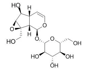
| Kinase Assay: |
| Int J Clin Exp Med. 2015 Feb 15;8(2):2038-44. | | Catalpol protects mice against renal ischemia/reperfusion injury via suppressing PI3K/Akt-eNOS signaling and inflammation.[Pubmed: 25932134] | Renal ischemia/reperfusion-injury (IRI) is a common disease in clinic, which is also the most common cause of acute kidney failure. Previous investigations has illustrated that Catalpol has neuroprotective, anti-inflammatory, and anti-hepatitis virus effects. This study was designed to investigate the protective effect of Catalpol on renal IRI mice through suppressing phosphatidylinositol 3-kinase/protein kinase B (PI3K/Akt)-endothelial nitric oxide synthase (eNOS) and against inflammation, and the possible underlying mechanism.
METHODS AND RESULTS:
Firstly, we used renal IRI model to analyze blood urea nitrogen and serum creatinine levels in renal IRI mice. Next, real-time PCR and western blotting were used to detect the expression of KIM-1 and the expression of PI3K, Akt and eNOS levels in renal IRI, respectively. In addition, activities of tumor necrosis factor α (TNF-α), interleukin-1β (IL-1β), interleukin-6 (IL-6) and interleukin-10 (IL-10) in renal IRI mice were measured with respective TNF-α, IL-1β, IL-6 and IL-10 ELISA kits. Our results showed that Catalpol clearly reduced blood urea nitrogen, serum creatinine levels and the expression of KIM-1 in renal IRI mice. Meanwhile, we found that Catalpol markedly reduced the expression of PI3K, Akt and eNOS levels in renal IRI group. Suppressing of the PI3K/Akt-eNOS and the TNF-α, IL-1β, IL-6 and IL-10 activities was involved in the protective effect of Catalpol on renal IRI.
CONCLUSIONS:
Collectively, Catalpol protected renal IRI via inhibiting PI3K/Akt-eNOS signaling and inflammatory responses. |
|
| Cell Research: |
| Biomed Pharmacother. 2015 Feb;69:291-6. | | Catalpol regulates cholinergic nerve system function through effect on choline acetyl-transferase not M receptor affinity.[Pubmed: 25661372] | To explore the effect of Catalpol on choline acetyl-transferase and M receptor affinity in a PC12 cell model and a rat model induced by beta-amyloid 25-35 (Aβ25-35).
METHODS AND RESULTS:
In PC12 cells, Catalpol (10μmol/l, 100μmol/) or saline was retained in the medium and Aβ25-35 (final concentration 20μmol/l) was added. Choline acetyl-transferase (ChAT) expression was determined by immunocytochemistry, ChAT activity measured by radioenzymatic assay, and M receptor (muscarinic receptor) affinity determined by (3)H-QNB binding test. In Wistar rats, Aβ25-35 was injected intracerebroventricularly to establish AD model. After injection of Aβ25-35, the rats were injected Catalpol at 5 and 10mg/kgd(-1) intraperitoneally for the next 7 days, and saline for the control rats. ChAT expression, ChAT activity and M receptor affinity were tested. Cells and rats all were divided into four groups: Group A (control), Group B (model), Group C (Catalpol low dose), and Group D (Catalpol high dose).
Compared with control, both PC12 cell and rat AD models showed decreased expression and activity of ChAT (p<0.01), but M receptor affinity remained the same (p>0.05). Compared with model group, treatment of Catalpol increased expression and activity of ChAT of PC12 cell and rat AD model induced by Aβ25-35, p<0.05 or p<0.01 respectively. But there was no difference of M receptor affinity among the four groups (p>0.05). M receptor affinity remained the same as concentration of Catalpol increased gradually in atropine competition experiments (p>0.05).
CONCLUSIONS:
Catalpol could regulate the cholinergic nerve system function from its effect on ChAT and may have beneficial effect for treatment of AD, but had no effect on M receptor affinity. | | Biosci Rep. 2016 Jun 30;36(3). pii: e00348. | | Catalpol inhibits apoptosis in hydrogen peroxide-induced cardiac myocytes through a mitochondrial-dependent caspase pathway.[Pubmed: 27166426] | Catalpol, an iridoid glucoside, has been reported to inhibit apoptosis of neuron and endothelial cells. In the present study, we investigated the mechanism of Catalpol-mediated cardioprotection.
METHODS AND RESULTS:
The rat embryonic ventricular myocardial cell line (H9c2) cells were first incubated with Catalpol, and then exposed to hydrogen peroxide (H2O2). The concentration of malondialdehyde (MDA) and the activity of superoxide dismutase (SOD) were all determined by using commercially available kits. Apoptotic cells were assessed by Hoechst 33258 and Annexin V-fluorescein isothiocyanate binding assay. Synthesis of Bcl-2, Bax, cytochrome c and caspase-3 were analysed by real-time semiquantitative reverse transcription-PCR and Western blotting. We observed that apoptosis in H9c2 was associated with increased Bax, cytochrome c, caspase-3, decreased Bcl-2 activity after 24 h of H2O2 exposure. Catalpol pretreatment afforded a marked protection against the above H2O2-mediated cytotoxicity and apoptosis in H9c2 cells. Moreover, the Catalpol pretreatment led to a great reduction in H2O2-induced MDA release and increased SOD.
CONCLUSIONS:
These findings indicated for the first time that pretreatment of H9c2 cells with Catalpol can be against H2O2-induced apoptosis, and the protective effect of Catalpol involves the mitochondrial-dependent caspase pathway and is associated with increased Bcl-2 and decreased Bax expression. |
|


 Cell. 2018 Jan 11;172(1-2):249-261.e12. doi: 10.1016/j.cell.2017.12.019.IF=36.216(2019)
 Cell Metab. 2020 Mar 3;31(3):534-548.e5. doi: 10.1016/j.cmet.2020.01.002.IF=22.415(2019)
 Mol Cell. 2017 Nov 16;68(4):673-685.e6. doi: 10.1016/j.molcel.2017.10.022.IF=14.548(2019)