| In vitro: |
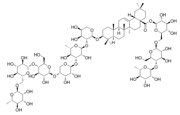
| Biomed Chromatogr. 2013 Dec;27(12):1767-74. | | Identification of the metabolites of anti-inflammatory compound clematichinenoside AR in rat intestinal microflora.[Pubmed: 23852993 ] | Clematichinenoside AR (C-AR), a pentacyclic triterpenoid saponin with anti-inflammatory and anti-rheumatoid activities, is the main active component of the traditional Chinese medicine Clematidis Radix et Rhizoma. However, its poor oral absorption indicated that not only the parent compound C-AR itself, but also its metabolites could be responsible for the pharmacological effects in rats. The present study aimed to investigate the metabolism of C-AR in rat intestinal microflora, where C-AR was extensively metabolized.
METHODS AND RESULTS:
C-AR was incubated with the content of the large intestine. The culture solution was collected at different time points and analyzed for the metabolites of C-AR. Eight metabolites were identified by liquid chromatography/quadrupole time-of-flight mass spectrometry. M1, M2 and M5 were the major metabolites. In addition, it was proposed that deglycosylation was the only pathway contributing to the biotransformation of C-AR in rat intestinal microflora. |
|
| In vivo: |
| Front Immunol. 2016 Dec 7;7:532. | | Succinate/NLRP3 Inflammasome Induces Synovial Fibroblast Activation: Therapeutical Effects of Clematichinenoside AR on Arthritis.[Pubmed: 28003810 ] | Clematichinenoside AR (C-AR) is a triterpene saponin isolated from the root of Clematis manshurica Rupr., which is a herbal medicine used in traditional Chinese medicine for the treatment of arthritis. C-AR exerts anti-inflammatory and immunosuppressive properties, but little is known about its action in the suppression of fibroblast activation. Low oxygen tension and transforming growth factor-β (TGF-β1) induction in the synovium contribute to fibrosis in arthritis. This study was designed to investigate the effect of C-AR on synovial fibrosis from the aspects of hypoxic TGF-β1 and hypoxia-inducible transcription factor-1α (HIF-1α) induction.
METHODS AND RESULTS:
In the synovium of rheumatoid arthritis (RA) rats, hypoxic TGF-β1 induction increased succinate accumulation due to the reversal of succinate dehydrogenase (SDH) activation and induced NLRP3 inflammasome activation in a manner dependent on HIF-1α induction. In response to NLRP3 inflammasome activation, the released IL-1β further increased TGF-β1 induction, suggesting the forward cycle between inflammation and fibrosis in myofibroblast activation. In the synovium of RA rats, C-AR inhibited hypoxic TGF-β1 induction and suppressed succinate-associated NLRP3 inflammasome activation by inhibiting SDH activity, and thereby prevented myofibroblast activation by blocking the cross-talk between inflammation and fibrosis.
CONCLUSIONS:
Taken together, these results showed that succinate worked as a metabolic signaling, linking inflammation with fibrosis through NLRP3 inflammasome activation. These findings suggested that synovial succinate accumulation and HIF-1α induction might be therapeutical targets for the prevention of fibrosis in arthritis. | | J Ethnopharmacol. 2014 Sep 11;155(2):1306-14. | | Clematichinenoside AR induces immunosuppression involving Treg cells in Peyer׳s patches of rats with adjuvant induced arthritis.[Pubmed: 25063305 ] | Clematichinenoside AR (AR) has been defined as a major active ingredient of triterpenoid saponins extracted from Clematidis Radix et Rhizoma, which is a traditional Chinese herbal medicine that has long been used in the treatment of rheumatoid arthritis (RA). To further explore the mechanism of AR in the treatment of RA, we investigated whether its immunomodulatory effects are related to Treg-mediated suppression derived from Peyer׳s patches (PPs) in adjuvant induced arthritis (AIA) rat model.
METHODS AND RESULTS:
AR (8, 16, 32 mg/kg) was orally administered daily from Day 18 to Day 31 after immunization. The effect of AR on AIA rats was evaluated by hind paw swelling and histopathological examination. Percentages of CD4(+)CD25(+)Foxp3(+) T regulatory cells were determined by flow cytometry. Levels of IL-10, TGF-β1, IL-17A and TNF-α were measured by ELISA. Expressions of Foxp3 and RORγ in synovium were detected using immunohistochemical analysis. AR treatment significantly reduced paw swelling of AIA rats, and histopathological analysis confirmed it could suppress severity of established arthritis. AR treatment upregulated the percentages of CD4(+)CD25(+)Foxp3(+) Treg cells among CD4+ T cells in PPs lymphocytes, and increased the levels of IL-10 and TGF-β1 secreted from ConA-activated PPs lymphocytes, whereas decreased the levels of IL-17 A and TNF-α. Similar tendency of circulating CD4(+)CD25(+)Foxp3(+) Treg cells percentages and serum cytokine levels were observed. Moreover, AR decreased the expression levels of Foxp3 and RORγ in joint synovial membrane.
CONCLUSIONS:
In conclusion, these results suggested AR has a potent protective effect on the progression of AIA, probably by augmenting CD4(+)CD25(+)Foxp3(+) Treg cells in PPs to induce immunosuppression, and modulating the balance between Treg cells and Th17 cells systemically. These findings may help to develop AR as a potent immunosuppressive agent for the treatment of RA. |
|


 Cell. 2018 Jan 11;172(1-2):249-261.e12. doi: 10.1016/j.cell.2017.12.019.IF=36.216(2019)
 Cell Metab. 2020 Mar 3;31(3):534-548.e5. doi: 10.1016/j.cmet.2020.01.002.IF=22.415(2019)
 Mol Cell. 2017 Nov 16;68(4):673-685.e6. doi: 10.1016/j.molcel.2017.10.022.IF=14.548(2019)