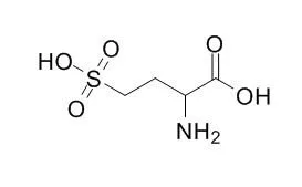
| In vivo: |
| Am J Physiol. 1989 Mar;256(3 Pt 2):H688-96. | | Cardiorespiratory effects of DL-homocysteic acid in caudal ventrolateral medulla.[Pubmed: 2646953] |
METHODS AND RESULTS:
We carried out experiments in urethan-anesthetized rats to determine 1) whether increasing the activity of small groups of neurons in the caudal ventrolateral medulla (CVLM) by injecting picomoles of an excitatory amino acid altered cardiovascular and/or respiratory homeostasis and 2) whether the depressor responses after chemical excitation in the CVLM were elicited only from the immunohistochemically identified catecholamine-containing cell group. In discrete sites in the CVLM, unilateral injections of 1-12 nl (20-240 pmol) of DL-Homocysteic acid (DLH; 20 mM, pH 7.4) selectively or concomitantly inhibited arterial pressure, heart rate, and diaphragm electromyogram (EMG) activity. In the region in which chemical excitation slowed breathing, units were recorded extracellularly that discharged with respiratory periodicity. Sites where the smallest volumes of DLH decreased arterial pressure were located outside the immunohistochemically identified DBH-positive cell bodies.
CONCLUSIONS:
These data suggest that either the same or neighboring neurons in the CVLM are involved in the central neural circuitry for both cardiovascular and respiratory control and that cells other than the catecholaminergic cell group are important in medullary depressor responses. | | Acta Neuropathol. 2002 May;103(5):428-36. | | DL-Homocysteic acid application disrupts calcium homeostasis and induces degeneration of spinal motor neurons in vivo.[Pubmed: 11935257] | Excitotoxicity, autoimmunity and free radicals have been postulated to play a role in the pathomechanism of amyotrophic lateral sclerosis (ALS), the most frequent motor neuron disease. Altered calcium homeostasis has already been demonstrated in Cu/Zn superoxide dismutase transgenic animals, suggesting a role for free radicals in the pathogenesis of ALS, and in passive transfer experiments, modeling autoimmunity. These findings also suggested that yet-confined pathogenic insults, associated with ALS, could trigger the disruption of calcium homeostasis of motor neurons.
METHODS AND RESULTS:
To test the possibility that excitotoxic processes may also be able to increase calcium in motor neurons, we applied the glutamate analogue DL-Homocysteic acid to the spinal cord of rats in vivo and analyzed the calcium distribution of the motor neurons over a 24-h survival period by electron microscopy. Initially, an elevated cytoplasmic calcium level, with no morphological sign of degeneration, was noticed. Later, increasing calcium accumulation was seen in different cellular compartments with characteristic features of alteration at different survival times. This calcium accumulation in organelles was paralleled by their progressive degeneration, which culminated in cell death by the end of the observation time. These findings confirm that increased calcium also plays a role in excitotoxic lesion of motor neurons, in line with previous studies documenting the involvement of calcium ions in motor neuronal injury in other models of the disease as well as elevated calcium in biopsy samples from ALS patients.
CONCLUSIONS:
We suggest that intracellular calcium might be responsible for the interplay between the different pathogenic processes resulting in a uniform clinicopathological picture of the disease.
|
|


 Cell. 2018 Jan 11;172(1-2):249-261.e12. doi: 10.1016/j.cell.2017.12.019.IF=36.216(2019)
 Cell Metab. 2020 Mar 3;31(3):534-548.e5. doi: 10.1016/j.cmet.2020.01.002.IF=22.415(2019)
 Mol Cell. 2017 Nov 16;68(4):673-685.e6. doi: 10.1016/j.molcel.2017.10.022.IF=14.548(2019)