| In vitro: |
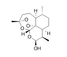
| PLoS One. 2015 Mar 23;10(3):e0120426. | | Dihydroartemisinin inhibits glucose uptake and cooperates with glycolysis inhibitor to induce apoptosis in non-small cell lung carcinoma cells.[Pubmed: 25799586] | Despite recent advances in the therapy of non-small cell lung cancer (NSCLC), the chemotherapy efficacy against NSCLC is still unsatisfactory. Previous studies show the herbal antimalarial drug Dihydroartemisinin (DHA) displays cytotoxic to multiple human tumors.
METHODS AND RESULTS:
Here, we showed that DHA decreased cell viability and colony formation, induced apoptosis in A549 and PC-9 cells. Additionally, we first revealed DHA inhibited glucose uptake in NSCLC cells. Moreover, glycolytic metabolism was attenuated by DHA, including inhibition of ATP and lactate production. Consequently, we demonstrated that the phosphorylated forms of both S6 ribosomal protein and mechanistic target of rapamycin (mTOR), and GLUT1 levels were abrogated by DHA treatment in NSCLC cells. Furthermore, the upregulation of mTOR activation by high expressed Rheb increased the level of glycolytic metabolism and cell viability inhibited by DHA. These results suggested that DHA-suppressed glycolytic metabolism might be associated with mTOR activation and GLUT1 expression. Besides, we showed GLUT1 overexpression significantly attenuated DHA-triggered NSCLC cells apoptosis. Notably, DHA synergized with 2-Deoxy-D-glucose (2DG, a glycolysis inhibitor) to reduce cell viability and increase cell apoptosis in A549 and PC-9 cells. However, the combination of the two compounds displayed minimal toxicity to WI-38 cells, a normal lung fibroblast cell line. More importantly, 2DG synergistically potentiated DHA-induced activation of caspase-9, -8 and -3, as well as the levels of both cytochrome c and AIF of cytoplasm. However, 2DG failed to increase the reactive oxygen species (ROS) levels elicited by DHA.
CONCLUSIONS:
Overall, the data shown above indicated DHA plus 2DG induced apoptosis was involved in both extrinsic and intrinsic apoptosis pathways in NSCLC cells. | | Cancer Res. 2005 Dec 1;65(23):10854-61. | | Dihydroartemisinin is cytotoxic to papillomavirus-expressing epithelial cells in vitro and in vivo.[Pubmed: 16322232 ] | Nearly all cervical cancers are etiologically attributable to human papillomavirus (HPV) infection and pharmaceutical treatments targeting HPV-infected cells would be of great medical benefit. Because many neoplastic cells (including cervical cancer cells) overexpress the transferrin receptor to increase their iron uptake, we hypothesized that iron-dependent, antimalarial drugs such as artemisinin might prove useful in treating HPV-infected or transformed cells.
METHODS AND RESULTS:
We tested three different artemisinin compounds and found that Dihydroartemisinin (DHA) and artesunate displayed strong cytotoxic effects on HPV-immortalized and transformed cervical cells in vitro with little effect on normal cervical epithelial cells. DHA-induced cell death involved activation of the mitochondrial caspase pathway with resultant apoptosis. Apoptosis was p53 independent and was not the consequence of drug-induced reductions in viral oncogene expression. Due to its selective cytotoxicity, hydrophobicity, and known ability to penetrate epithelial surfaces, we postulated that DHA might be useful for the topical treatment of mucosal papillomavirus lesions. To test this hypothesis, we applied DHA to the oral mucosa of dogs that had been challenged with the canine oral papillomavirus. Although applied only intermittently, DHA strongly inhibited viral-induced tumor formation. Interestingly, the DHA-treated, tumor-negative dogs developed antibodies against the viral L1 capsid protein, suggesting that DHA had inhibited tumor growth but not early rounds of papillomavirus replication.
CONCLUSIONS:
These findings indicate that DHA and other artemisinin derivatives may be useful for the topical treatment of epithelial papillomavirus lesions, including those that have progressed to the neoplastic state. |
|


 Cell. 2018 Jan 11;172(1-2):249-261.e12. doi: 10.1016/j.cell.2017.12.019.IF=36.216(2019)
 Cell Metab. 2020 Mar 3;31(3):534-548.e5. doi: 10.1016/j.cmet.2020.01.002.IF=22.415(2019)
 Mol Cell. 2017 Nov 16;68(4):673-685.e6. doi: 10.1016/j.molcel.2017.10.022.IF=14.548(2019)