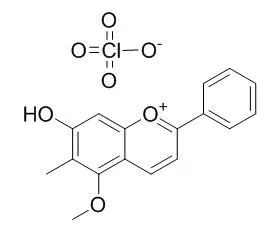
| In vitro: |
| Eur J Pharmacol. 2014 Apr 5;728:82-92. | | Dracorhodin perchlorate induces apoptosis in primary fibroblasts from human skin hypertrophic scars via participation of caspase-3.[Pubmed: 24525335] | Hypertrophic scar (HS) is an abnormally proliferative disorder characterized by excessive proliferation of fibroblasts and redundant deposition of extracellular matrix. An unbalance between fibroblast proliferation and apoptosis has been assumed to play an important role in HS formation.
METHODS AND RESULTS:
To explore the regulative effects of Dracorhodin perchlorate (Dp), one of the derivants of dracorhodin that is a major constituent in the traditional Chinese medicine, on primary fibroblasts from human skin hypertrophic scars, 3-[4,5-dimethylthiazol-2-yl]-2,5-diphenyltetrazolium bromide (MTT) assay and flow cytometric analysis were respectively used to evaluate the inhibitory effect of Dp on the cells and to determine cell cycle distribution. Additionally, cellular apoptosis was separately detected with Hoechst 33258 staining and terminal deoxynucleotidyl transferase (TdT)-mediated dUTP nick end labeling (TUNEL) assay. The expression levels of caspase-3 mRNA and protein were respectively measured with reverse transcription-polymerase chain reaction and western blot analysis, and caspase-3 activity were determined using a colorimetric assay kit. The results showed that Dp significantly inhibited cell growth, and induced apoptosis in fibroblasts in a dose-and time-dependent manner, arresting cell cycle at G1 phase. Additionally, Dp slightly up-regulated caspase-3 mRNA expression in fibroblasts, but significantly down-regulated caspase-3 protein expression in a dose- and time-dependent manner, and concurrently elevated caspase-3 activity.
CONCLUSIONS:
Taken together, these data indicated that Dp could effectively inhibit cell proliferation, and induced cell cycle arrest and apoptosis in fibroblasts, at least partially via modulation of caspase-3 expression and its activity, which suggests that Dp is an effective and potential candidate to develop for HS treatment. | | Zhongguo Zhong Yao Za Zhi. 2010 Aug;35(15):1996-2000. | | Dracorhodin perchlorate inhibit high glucose induce serum and glucocorticoid induced protein kinase 1 and fibronectin expression in human mesangial cells.[Pubmed: 20931854] | To investigate the effect of Dracorhodin perchlorate (DP) on inhibiting high glucose-induced serum and glucocorticoid induced protein kinase 1 (SGK1) and fibronectin (FN) expression in human mesangial cells (HMC), and its mechanism of prevention and treatment on renal fibrosis in diabetic nephropathy (DN) .
METHODS AND RESULTS:
The HMC were divided into normal glucose group (NG group, 5.5 mmol x L(-1) D-glucose), normal glucose +low DP group (NG + LDP group, 5.5 mmol x L(-1) D-glucose +7.5 micromol x L(-1) DP), normal glucose +high DP group (NG + HDP group, 5.5 mmol x L(-1) D-glucose + 15 micromol x L(-1) DP), high glucose group (HG group,25 mmol x L(-1) D-glucose), high glucose +low DP group (HG + LDP group, 25 mmol x L(-1) D-glucose + 7.5 micromol x L(-1) DP)and high glucose +high DP group (HG +HDP group, 25 mmol x L(-1) D-glucose + 15 micromol x L(-1) DP). Each group was examined at 24 hours. The levels of SGK1 and FN mRNA was detected by real-time fluorescence quantitative PCR,and the expression of SGK1 and FN protein was detected by Western blot or indirect immunofluorescence.
A basal level of SGK1 and FN in HMC were detected in NG group, and the level of SGK1 and FN mRNA and protein were not evidently different compared to that of NG group adding 7.5 micromol x L(-1) DP for 24 hours. On the other hand, the levels of SGK1 and FN mRNA and protein were obviously decreased by adding 15 micromol x L(-1) DP for 24 hours. Compared to NG group, the levels of SGK1 and FN mRNA and protein were increased in HG group after stimulating for 24 hours (P < 0.01). Compared to HG group, the level of SGK1 and FN mRNA and protein were evidently reduced in HG + LDP and HG + HDP groups by adding 7.5 micromol x L(-1) DP and 15 micromol x L(-1) DP for 24 hours (P < 0.01).
CONCLUSIONS:
Dracorhodin perchlorate can inhibit high glucose-induced serum and glucocorticoid induced protein kinase 1 (SGK1) and fibronectin(FN) expression in human mesangial cells, and this may be part of the mechanism of preventing and treating renal fibrosis of DN. | | J Huazhong Univ Sci Technolog Med Sci. 2011 Apr;31(2):215-9. | | Dracorhodin perchlorate suppresses proliferation and induces apoptosis in human prostate cancer cell line PC-3.[Pubmed: 21505988] |
METHODS AND RESULTS:
The growth inhibition and pro-apoptosis effects of Dracorhodin perchlorate on human prostate cancer PC-3 cell line were examined. After administration of 10-80 μmol/L Dracorhodin perchlorate for 12-48 h, cell viability of PC-3 cells was measured by MTT colorimetry. Cell proliferation ability was detected by colony formation assay. Cellular apoptosis was inspected by acridine orange-ethidium bromide fluorescent staining, Hoechst 33258 fluorescent staining, and flow cytometry (FCM) with annexin V-FITC/propidium iodide dual staining. The results showed that Dracorhodin perchlorate inhibited the growth of PC-3 in a dose- and time-dependent manner. IC50 of Dracorhodin perchlorate on PC-3 cells at 24 h was 40.18 μmol/L. Cell clone formation rate was decreased by 86% after treatment with 20 μmol/L of Dracorhodin perchlorate. Some cells presented the characteristic apoptotic changes. The cellular apoptotic rates induced by 10-40 μmol/L Dracorhodin perchlorate for 24 h were 8.43% to 47.71% respectively.
CONCLUSIONS:
It was concluded that Dracorhodin perchlorate significantly inhibited the growth of PC-3 cells by suppressing proliferation and inducing apoptosis of the cells. |
|


 Cell. 2018 Jan 11;172(1-2):249-261.e12. doi: 10.1016/j.cell.2017.12.019.IF=36.216(2019)
 Cell Metab. 2020 Mar 3;31(3):534-548.e5. doi: 10.1016/j.cmet.2020.01.002.IF=22.415(2019)
 Mol Cell. 2017 Nov 16;68(4):673-685.e6. doi: 10.1016/j.molcel.2017.10.022.IF=14.548(2019)