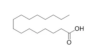
| In vitro: |
| Diabetes. 2001 Sep;50(9):2105-13. | | Glucose and palmitic acid induce degeneration of myofibrils and modulate apoptosis in rat adult cardiomyocytes.[Pubmed: 11522678] | Several studies support the concept of a diabetic cardiomyopathy in the absence of discernible coronary artery disease, although its mechanism remains poorly understood. We investigated the role of glucose and Palmitic acid on cardiomyocyte apoptosis and on the organization of the contractile apparatus.
METHODS AND RESULTS:
Exposure of adult rat cardiomyocytes for 18 h to Palmitic acid (0.25 and 0.5 mmol/l) resulted in a significant increase of apoptotic cells, whereas increasing glucose concentration to 33.3 mmol/l for up to 8 days had no influence on the apoptosis rate. However, both Palmitic acid and elevated glucose concentration alone or in combination had a dramatic destructive effect on the myofibrillar apparatus. The membrane-permeable C2-ceramide but not the metabolically inactive C2-dihydroceramide enhanced apoptosis of cardiomyocytes by 50%, accompanied by detrimental effects on the myofibrils. The Palmitic acid-induced effects were impaired by fumonisin B1, an inhibitor of ceramide synthase. Sphingomyelinase, which activates the catabolic pathway of ceramide by metabolizing sphingomyeline to ceramide, did not adversely affect cardiomyocytes. Palmitic acid-induced apoptosis was accompanied by release of cytochrome c from the mitochondria. Aminoguanidine did not prevent glucose-induced myofibrillar degeneration, suggesting that formation of nitric oxide and/or advanced glycation end products play no major role.
CONCLUSIONS:
Taken together, these results suggest that in adult rat cardiac cells, Palmitic acid induces apoptosis via de novo ceramide formation and activation of the apoptotic mitochondrial pathway. Conversely, glucose has no influence on adult cardiomyocyte apoptosis. However, both cell nutrients promote degeneration of myofibrils. Thus, gluco- and lipotoxicity may play a central role in the development of diabetic cardiomyopathy. | | Anticancer Res. 2002 Sep-Oct;22(5):2587-90. | | Antitumor activity of palmitic acid found as a selective cytotoxic substance in a marine red alga.[Pubmed: 12529968] | In a previous report, we discussed an extract from a marine red alga, Amphiroa zonata, which shows selective cytotoxic activity to human leukemic cells, but no cytotoxicity to normal human dermal fibroblast (HDF) cells in vitro.
METHODS AND RESULTS:
In this study, we identified Palmitic acid, a selective cytotoxic substance from the marine algal extract, and investigated its biological activities. At concentrations ranging from 12.5 to 50 micrograms/ml, Palmitic acid shows selective cytotoxicity to human leukemic cells, but no cytotoxicity to normal HDF cells. Furthermore, Palmitic acid induces apoptosis in the human leukemic cell line MOLT-4 at 50 micrograms/ml. Palmitic acid also shows in vivo antitumor activity in mice. One molecular target of Palmitic acid in tumor cells is DNA topoisomerase I, however, interestingly, it does not affect DNA topoisomerase II,
CONCLUSIONS:
suggesting that Palmitic acid may be a lead compound of anticancer drugs. |
|
| In vivo: |
| Metabolism. 2014 Sep;63(9):1131-40. | | The saturated fatty acid, palmitic acid, induces anxiety-like behavior in mice.[Pubmed: 25016520] | Excess fat in the diet can impact neuropsychiatric functions by negatively affecting cognition, mood and anxiety. We sought to show that the free fatty acid (FFA), Palmitic acid, can cause adverse biobehaviors in mice that last beyond an acute elevation in plasma FFAs.
METHODS AND RESULTS:
Mice were administered Palmitic acid or vehicle as a single intraperitoneal (IP) injection. Biobehaviors were profiled 2 and 24 h after Palmitic acid treatment. Quantification of dopamine (DA), norepinephrine (NE), serotonin (5-HT) and their major metabolites was performed in cortex, hippocampus and amygdala. FFA concentration was determined in plasma. Relative fold change in mRNA expression of unfolded protein response (UPR)-associated genes was determined in brain regions.
In a dose-dependent fashion, Palmitic acid rapidly reduced mouse locomotor activity by a mechanism that did not rely on TLR4, MyD88, IL-1, IL-6 or TNFα but was dependent on fatty acid chain length. Twenty-four hours after Palmitic acid administration mice exhibited anxiety-like behavior without impairment in locomotion, food intake, depressive-like behavior or spatial memory. Additionally, the serotonin metabolite 5-HIAA was increased by 33% in the amygdala 24h after Palmitic acid treatment.
CONCLUSIONS:
Palmitic acid induces anxiety-like behavior in mice while increasing amygdala-based serotonin metabolism. These effects occur at a time point when plasma FFA levels are no longer elevated. |
|


 Cell. 2018 Jan 11;172(1-2):249-261.e12. doi: 10.1016/j.cell.2017.12.019.IF=36.216(2019)
 Cell Metab. 2020 Mar 3;31(3):534-548.e5. doi: 10.1016/j.cmet.2020.01.002.IF=22.415(2019)
 Mol Cell. 2017 Nov 16;68(4):673-685.e6. doi: 10.1016/j.molcel.2017.10.022.IF=14.548(2019)