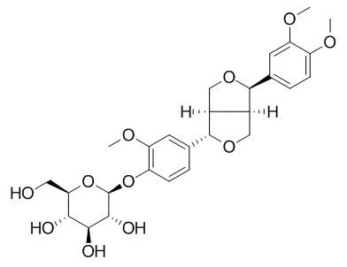
| Cell Research: |
| Cell Mol Neurobiol. 2014 Nov;34(8):1165-73. | | Protective effects of phillyrin on H2O 2-induced oxidative stress and apoptosis in PC12 cells.[Pubmed: 25146667] | Oxidative stress is a major component of harmful cascades activated in neurodegenerative disorders. We sought to elucidate possible effects of Phillyrin, an active constituent isolated from the Chinese medicinal herb Forsythia suspense, on hydrogen peroxide (H2O2)-induced cell death and determine the underlying molecular mechanisms in neuron-like PC12 cells.
METHODS AND RESULTS:
By MTT assay and lactate dehydrogenase (LDH) leakage assay, we found that Phillyrin treatment effectively protected PC12 cells against H2O2-induced cell damage. H2O2 exposure induced oxidative stress in PC12 cells, as revealed by enhanced oxidative stress and decreased activities of antioxidative enzymes, which were inhibited by Phillyrin pretreatment. ROS activated mitochondria-dependent apoptosis. The anti-apoptotic effects of Phillyrin were also confirmed by acridine orange/ethidium bromide (AO/EB) staining. Mitochondrial membrane potential decrease, cytochrome c release, caspases activation, activation of AIF and Endo G were observed in H2O2-treated cells by rhodamine 123 or western blot. Interestingly, Phillyrin effectively suppressed these changes. Moreover, Phillyrin could inhibit H2O2-induced up-regulation of Bax/Bcl-2 ratio.
CONCLUSIONS:
In conclusion, Phillyrin effectively inhibited H2O2-induced oxidative stress and apoptosis in PC12 cells. |
|
| Animal Research: |
| Eur J Drug Metab Pharmacokinet. 2009 Apr-Jun;34(2):79-83. | | Pharmacokinetics of phillyrin and forsythiaside following iv administration to Beagle dog.[Pubmed: 19645216] | The objective of the present study was to firstly investigate the in vivo pharmacokinetics of Phillyrin and forsythiaside in beagle dog.
METHODS AND RESULTS:
On I.V. administration, a rapid distribution was observed and followed by a slower elimination for Phillyrin and forsythiaside. The mean t(1/2Z) was 49.99, 34.87 and 43.81 min for 0.19, 0.70 and 1.43 mg/kg of Phillyrin, and 60.90, 64.30, 57.99 min for 0.62, 1.39 and 5.52 mg/kg of forsythiaside respectively. And the AUC(o-t) increased linearly from 36.51 to 160.22 microg x min/ml of Phillyrin and from 50.63 to 681.08 microg x min/ml after the three dosage administrated. In the range of the dose examined, the pharmacokinetics of Phillyrin and forsythiaside in beagle dog was based on first order kinetics.
CONCLUSIONS:
Although both drugs were widely distributed to various tissues in the dog, no concerns about extensive binding to tissues that may be consumed by the public should a dog be exposed to Phillyrin and forsythiaside according to the rapid elimination. | | Fitoterapia. 2013 Oct;90:132-9. | | Phillyrin attenuates LPS-induced pulmonary inflammation via suppression of MAPK and NF-κB activation in acute lung injury mice.[Pubmed: 23751215] | Phillyrin (Phil) is one of the main chemical constituents of Forsythia suspensa (Thunb.), which has shown to be an important traditional Chinese medicine. We tested the hypothesis that Phil modulates pulmonary inflammation in an ALI model induced by LPS.
METHODS AND RESULTS:
Male BALB/c mice were pretreated with or without Phil before respiratory administration with LPS, and pretreated with dexamethasone as a control. Cytokine release (TNF-α, IL-1β, and IL-6) and amounts of inflammatory cell in bronchoalveolar lavage fluid (BALF) were detected by ELISA and cell counting separately. Pathologic changes, including neutrophil infiltration, interstitial edema, hemorrhage, hyaline membrane formation, necrosis, and congestion during acute lung injury in mice were evaluated via pathological section with HE staining. To further investigate the mechanism of Phil anti-inflammatory effects, activation of MAPK and NF-κB pathways was tested by western blot assay. Phil pretreatment significantly attenuated LPS-induced pulmonary histopathologic changes, alveolar hemorrhage, and neutrophil infiltration. The lung wet-to-dry weight ratios, as the index of pulmonary edema, were markedly decreased by Phil pretreatment. In addition, Phil decreased the production of the proinflammatory cytokines including (TNF-α, IL-1β, and IL-6) and the concentration of myeloperoxidase (MPO) in lung tissues. Phil pretreatment also significantly suppressed LPS-induced activation of MAPK and NF-κB pathways in lung tissues.
CONCLUSIONS:
Taken together, the results suggest that Phil may have a protective effect on LPS-induced ALI, and it potentially contributes to the suppression of the activation of MAPK and NF-κB pathways. Phil may be a new preventive agent of ALI in the clinical setting. |
|


 Cell. 2018 Jan 11;172(1-2):249-261.e12. doi: 10.1016/j.cell.2017.12.019.IF=36.216(2019)
 Cell Metab. 2020 Mar 3;31(3):534-548.e5. doi: 10.1016/j.cmet.2020.01.002.IF=22.415(2019)
 Mol Cell. 2017 Nov 16;68(4):673-685.e6. doi: 10.1016/j.molcel.2017.10.022.IF=14.548(2019)