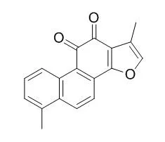
| Cell Research: |
| Food Chem Toxicol. 2013 Dec;62:407-12. | | Protective properties of tanshinone I against oxidative DNA damage and cytotoxicity.[Pubmed: 24021569] | Tanshinone I, a naturally occurring diterpene from Danshen, has been shown to possess hepatocyte protective, anticancer, and memory enhancing properties. However, there are few stringent pharmacological tests for neuroprotection of Tanshinone I thus far. Since peroxynitrite is involved in the pathogenesis of neurodegenerative disorders, this study was undertaken to investigate whether the neuroprotective effect of Tanshinone I is associated with inhibition of peroxynitrite-caused DNA damage, a critical event leading to peroxynitrite-induced cytotoxicity.
METHODS AND RESULTS:
Our results show that Tanshinone I can significantly inhibit peroxynitrite-induced DNA damage both in φX-174 plasmid DNA and rat primary astrocytes. EPR spectroscopy indicates that Tanshinone I potently diminished the DMPO-hydroxyl radical adduct signal from peroxynitrite.
CONCLUSIONS:
Taken together, these results demonstrate for the first time that Tanshinone I can protect against peroxynitrite-induced DNA damage, hydroxyl radical formation and cytotoxicity, which might have implications for Tanshinone I-mediated neuroprotection. | | Redox Biol. 2013 Oct 29;1:532-41. | | The Nrf2-inducers tanshinone I and dihydrotanshinone protect human skin cells and reconstructed human skin against solar simulated UV.[Pubmed: 24273736] | Exposure to solar ultraviolet (UV) radiation is a causative factor in skin photocarcinogenesis and photoaging, and an urgent need exists for improved strategies for skin photoprotection. The redox-sensitive transcription factor Nrf2 (nuclear factor-E2-related factor 2), a master regulator of the cellular antioxidant defense against environmental electrophilic insult, has recently emerged as an important determinant of cutaneous damage from solar UV, and the concept of pharmacological activation of Nrf2 has attracted considerable attention as a novel approach to skin photoprotection. In this study, we examined feasibility of using tanshinones, a novel class of phenanthrenequinone-based cytoprotective Nrf2 inducers derived from the medicinal plant Salvia miltiorrhiza, for protection of cultured human skin cells and reconstructed human skin against solar simulated UV.
METHODS AND RESULTS:
Using a dual luciferase reporter assay in human Hs27 dermal fibroblasts pronounced transcriptional activation of Nrf2 by four major tanshinones [Tanshinone I (T-I), dihydrotanshinone (DHT), Tanshinone IIA (T-II-A) and cryptotanshinone (CT)] was detected. In fibroblasts, the more potent tanshinones T-I and DHT caused a significant increase in Nrf2 protein half-life via blockage of ubiquitination, ultimately resulting in upregulated expression of cytoprotective Nrf2 target genes (GCLC, NQO1) with the elevation of cellular glutathione levels. Similar tanshinone-induced changes were also observed in HaCaT keratinocytes. T-I and DHT pretreatment caused significant suppression of skin cell death induced by solar simulated UV and riboflavin-sensitized UVA. Moreover, feasibility of tanshinone-based cutaneous photoprotection was tested employing a human skin reconstruct exposed to solar simulated UV (80 mJ/cm(2) UVB; 1.53 J/cm(2) UVA). The occurrence of markers of epidermal solar insult (cleaved procaspase 3, pycnotic nuclei, eosinophilic cytoplasm, acellular cavities) was significantly attenuated in DHT-treated reconstructs that displayed increased immunohistochemical staining for Nrf2 and γ-GCS together with the elevation of total glutathione levels.
CONCLUSIONS:
Taken together, our data suggest the feasibility of achieving tanshinone-based cutaneous Nrf2-activation and photoprotection. | | Int J Mol Med. 2008 Nov;22(5):613-8. | | Growth inhibition and apoptosis induction by tanshinone I in human colon cancer Colo 205 cells.[Pubmed: 18949381] | Tanshinone I (Tan-I) and Tanshinone IIA (Tan-IIA) were isolated from Danshen (Salviae Miltiorrhizae Radix), a widely prescribed traditional herbal medicine that is used to treat cardiovascular and dysmenorrhea diseases.
In our previous study, Tan-IIA was demonstrated to induce apoptosis in human colon cancer Colo 205 cells. However, the effect of Tan-I on human colon cancer cells is not clearly understood yet.
METHODS AND RESULTS:
In this study, the anti-growth and apoptosis-eliciting effects of Tan-I, as well as its cellular mechanisms of actions, were investigated in Colo 205 human colon cancer cells. Tan-I reduced cell growth in a concentration-dependent manner, inducing apoptosis accompanied by an increase in TUNEL staining and in cells in the sub-G1 fraction. The expression of p53, p21, bax and caspase-3 increased in Tan-I-treated cells. In addition, the cell cycle analysis showed G0/G1 arrest.
CONCLUSIONS:
These findings suggest that Tan-I induces apoptosis in Colo 205 cells through both mitochondrial-mediated intrinsic cell-death pathways and p21-mediated G0/G1cell cycle arrest. Accordingly, the therapeutic potential of Tan-I for colon cancer deserves further study. |
|
| Animal Research: |
| Neurochem. Res., 2016,41(8):1958-68. | | Tanshinone I Enhances Neurogenesis in the Mouse Hippocampal Dentate Gyrus via Increasing Wnt-3, Phosphorylated Glycogen Synthase Kinase-3β and β-Catenin Immunoreactivities.[Pubmed: 27053301 ] | Tanshinone I (TsI), a lipophilic diterpene extracted from Danshan (Radix Salvia miltiorrhizae), exerts neuroprotection in cerebrovascular diseases including transient ischemic attack.
In this study, we examined effects of TsI on cell proliferation and neuronal differentiation in the subgranular zone (SGZ) of the mouse dentate gyrus (DG) using Ki-67, BrdU and doublecortin (DCX) immunohistochemistry.
METHODS AND RESULTS:
Mice were treated with 1 and 2 mg/kg TsI for 28 days. In the 1 mg/kg TsI-treated-group, distribution patterns of BrdU, Ki-67 and DCX positive ((+)) cells in the SGZ were similar to those in the vehicle-treated-group. However, in the 2 mg/kg TsI-treated-group, double labeled BrdU(+)/NeuN(+) cells, which are mature neurons, as well as Ki-67(+), DCX(+) and BrdU(+) cells were significantly increased compared with those in the vehicle-treated-group. On the other hand, immunoreactivities and protein levels of Wnt-3, β-catenin and serine-9-glycogen synthase kinase-3β (p-GSK-3β), which are related with morphogenesis, were significantly increased in the granule cell layer of the DG only in the 2 mg/kg TsI-treated-group.
CONCLUSIONS:
Therefore, these findings indicate that TsI can promote neurogenesis in the mouse DG and that the neurogenesis is related with increases of Wnt-3, p-GSK-3β and β-catenin immunoreactivities. |
|


 Cell. 2018 Jan 11;172(1-2):249-261.e12. doi: 10.1016/j.cell.2017.12.019.IF=36.216(2019)
 Cell Metab. 2020 Mar 3;31(3):534-548.e5. doi: 10.1016/j.cmet.2020.01.002.IF=22.415(2019)
 Mol Cell. 2017 Nov 16;68(4):673-685.e6. doi: 10.1016/j.molcel.2017.10.022.IF=14.548(2019)