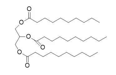
| Animal Research: |
| Journal of oleo ence, 2018, 67(8). | | Tricaprin Rescues Myocardial Abnormality in a Mouse Model of Triglyceride Deposit Cardiomyovasculopathy[Reference: WebLink] | Triglyceride deposit cardiomyovasculopathy (TGCV) is an intractable cardiovascular disease for which a specific treatment is urgently required. In TGCV, adipose triglyceride lipase (ATGL) deficiency results in the abnormal intracellular metabolism of long-chain fatty acid (LCFA) which leads to TG deposition. Medium-chain triglycerides have been used as an important functional food for various human diseases.
METHODS AND RESULTS:
To address the potential activities of Tricaprin, a medium-chain triglyceride, on cardiac dysfunctions of TGCV, we examined the effects of Tricaprin diet on Atgl knock out (KO) mice, an animal model for TGCV. Cardiac imaging tests showed that the Tricaprin diet reduced TG accumulation, resulting from improvement of LCFA metabolism, and improved left ventricular function in Atgl KO mice compared to that in mice fed the control diet.
CONCLUSIONS:
In conclusion, Tricaprin improved myocardial abnormality in the TGCV model, thus, it may be useful for the treatment of patients with TGCV. | | Aquaculture, 2000, 190(3-4):261-271. | | Growth and survival of European sea bass (Dicentrarchus Labrax) larvae fed from first feeding on compound diets containing medium-chain triacylglycerols.[Reference: WebLink] | A 21-day feeding trial was carried out to investigate the ability of first feeding European sea bass larvae to utilize medium-chain triacylglycerols as an alternative source of energy.
METHODS AND RESULTS:
Three compound diets based on soluble fish protein concentrate and yeast were supplemented with either 3% tricaproin (TC6), tricaprylin (TC8) or Tricaprin (TC10). A diet containing triolein (TOL) was used as a reference diet. Diets were tested on four replicate groups of first feeding European sea bass larvae at 20°C, i.e. 6 days after hatching. At the end of the 21-day trial, TC8 yielded significantly higher survival (57±8% vs. 28±11% for the three other groups). Considered together, larvae fed TC8 and TC6 displayed better growth rates than larvae fed TOL and TC10 (final mean wet weights: 1.5±0.3 mg vs. 1.2±0.2 mg, respectively). The fatty acid composition of larval total lipid revealed a low deposit of medium-chain fatty acids (between 1 and 3% of total fatty acids) suggesting that medium-chain fatty acids were oxidized for energetic purposes.
CONCLUSIONS:
Tricaprylin and to a lesser extent tricaproin, appear to be potential energy sources for first feeding European sea bass larvae reared on compound diets. |
|
| Structure Identification: |
| Biomaterials, 2002, 23(16):3465-3471. | | Effect of tricaprin on the physical characteristics and in vitro release of etoposide from PLGA microspheres.[Reference: WebLink] | The purpose of this article is to examine the effects of Tricaprin on the physical characteristics and in vitro release of etoposide from poly (lactic-co-glycolic acid) microspheres.
METHODS AND RESULTS:
The microspheres were synthesized through the use of a single-emulsion solvent-extraction procedure. Samples from each batch of microspheres were then analyzed for size distribution, drug loading efficiency, surface characteristics, in vitro release, and in vitro degradation of microspheres. Microsphere batches were synthesized using three different etoposide concentrations (15%, 10%, and 5% w/w) with Tricaprin concentrations of 25% and 50%. The incorporation of 50% Tricaprin significantly increased (p<0.05) the size of the microspheres for all three etoposide concentrations in comparison to microspheres prepared without Tricaprin (control). The percentage of Tricaprin used did not significantly affect the drug loading efficiency of the microspheres. The addition of Tricaprin was shown to significantly increase (p<0.05) the in vitro release of etoposide from the microspheres prepared with all three concentrations of etoposide and the two different Tricaprin percentages. Examination of the surface characteristics of the Tricaprin loaded microspheres showed a dimpled surface with what appeared to be pockets of Tricaprin dispersed throughout.
CONCLUSIONS:
In the in vitro degradation study, the Tricaprin microspheres grew very porous as the degradation time increased, but they still retained a recognizable structure even after 30 days of degradation. |
|


 Cell. 2018 Jan 11;172(1-2):249-261.e12. doi: 10.1016/j.cell.2017.12.019.IF=36.216(2019)
 Cell Metab. 2020 Mar 3;31(3):534-548.e5. doi: 10.1016/j.cmet.2020.01.002.IF=22.415(2019)
 Mol Cell. 2017 Nov 16;68(4):673-685.e6. doi: 10.1016/j.molcel.2017.10.022.IF=14.548(2019)