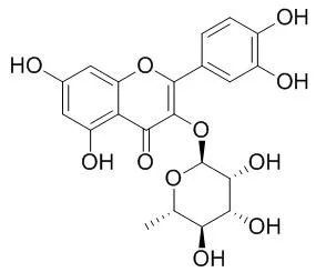
| Kinase Assay: |
| Biochem Pharmacol. 2005 Jun 15;69(12):1839-51. | | Quercetin, but not rutin and quercitrin, prevention of H2O2-induced apoptosis via anti-oxidant activity and heme oxygenase 1 gene expression in macrophages.[Pubmed: 15876423] | In the present study, we examine the protective mechanism of quercetin (QE) on oxidative stress-induced cytotoxic effect in RAW264.7 macrophages.
METHODS AND RESULTS:
Results of Western blotting show that QE but not its glycoside rutin (RUT) and quicitrin-induced HO-1 protein expression in a time- and dose-dependent manner, and HO-1 protein induced by QE was blocked by an addition of cycloheximide or actinomycin D. Induction of HO-1 gene expression by QE was accompanied by inducing ERKs, but not JNKs or p38, proteins phosphorylation. Addition of PD98059, but not SB203580 or SP600125, significantly attenuates QE-induced HO-1 protein and mRNA expression associated with blocking the expression of phosphorylated ERKs proteins. H(2)O(2) addition reduces the viability of cells by MTT assay, and appearance of DNA ladders, hypodiploid cells, and an increase in intracellular peroxide level was detected. Addition of QE, but not QI or RUT, significantly reduced the cytotoxic effect induced by H(2)O(2) associated with blocking the production of intracellular peroxide, DNA ladders, and hypodiploid cells. QE protection of cells from H(2)O(2)-induced apoptosis was significantly suppressed by adding HO inhibitor SnPP or ERKs inhibitor PD98059. Additionally, QE protects cells from H(2)O(2)-induced a decrease in the mitochondrial membrane potential and a release of cytochrome c from mitochondria to cytosol by DiOC6 and Western blotting assay, respectively. Activation of apoptotic proteins including the caspase 3, caspase 9, PARP, D4-GDI proteins was identified in H(2)O(2)-treated cells by Western blotting and enzyme activity assay, and that was significantly blocked by an addition of QE, but not RUT and QI. Furthermore, HO-1 catalytic metabolites carbon monoxide (CO), but not Fe(2+), Fe(3+), biliverdin or bilirubin, performed protective effect on cells from H(2)O(2)-induced cell death with an increase in HO-1 protein expression and ERKs protein phosphorylation.
CONCLUSIONS:
These data suggest that induction of HO-1 protein may participate in the protective mechanism of QE on oxidative stress (H(2)O(2))-induced apoptosis, and reduction of intracellular ROS production and mitochondria dysfunction with blocking apoptotic events were involved. Differential anti-apoptotic effect between QE and its glycosides RUT and QI via distinct HO-1 protein induction was also delineated. |
|
| Cell Research: |
| Pathol Oncol Res. 2015 Apr;21(2):333-8. | | Apoptotic Effects of Quercitrin on DLD-1 Colon Cancer Cell Line.[Pubmed: 25096395] | Quercetin, which is the most abundant bioflavonoid compound, is mainly present in the glycoside form of Quercitrin. Although different studies indicated that Quercitrin is a potent antioxidant, the action of this compound is not well understood. In this study, we investigated whether Quercitrin has apoptotic and antiproliferative effects in DLD-1 colon cancer cell lines.
METHODS AND RESULTS:
Time and dose dependent antiproliferative and apoptotic effects of Quercitrin were subsequently determined by WST-1 cell proliferation assay, lactate dehydrogenase (LDH) cytotoxicity assay, detection of nucleosome enrichment factor, changes in caspase-3 activity, loss of mitochondrial membrane potential (MMP) and also the localization of phosphatidylserine (PS) in the plasma membrane. There were significant increases in caspase-3 activity, loss of MMP, and increases in the apoptotic cell population in response to Quercitrin in DLD-1 colon cancer cells in a time- and dose-dependent manner.
CONCLUSIONS:
These results revealed that Quercitrin has antiproliferative and apoptotic effects on colon cancer cells. Quercitrin activity supported with in vivo analyses could be a biomarker candicate for early colorectal carcinoma. | | Biochem Pharmacol. 2013 Nov 15;86(10):1476-86. | | Quercitrin and taxifolin stimulate osteoblast differentiation in MC3T3-E1 cells and inhibit osteoclastogenesis in RAW 264.7 cells.[Pubmed: 24060614] | Flavonoids are natural antioxidants that positively influence bone metabolism. The present study screened among different flavonoids to identify biomolecules for potential use in bone regeneration. For this purpose, we used MC3T3-E1 and RAW264.7 cells to evaluate their effect on cell viability and cell differentiation.
METHODS AND RESULTS:
First, different doses of chrysin, diosmetin, galangin, Quercitrin and taxifolin were analyzed to determine the optimum concentration to induce osteoblast differentiation. After 48h of treatment, doses ≥100μM of diosmetin and galangin and also 500μM taxifolin revealed a toxic effect on cells. The same effect was observed in cells treated with doses ≥100μM of chrysin after 14 days of treatment. However, the safe doses of Quercitrin (200 and 500μM) and taxifolin (100 and 200μM) induced bone sialoprotein and osteocalcin mRNA expression. Also higher osteocalcin secreted levels were determined in 100μM taxifolin osteoblast treated samples when compared with the control ones. On the other hand, Quercitrin and taxifolin decreased Rankl gene expression in osteoblasts, suggesting an inhibition of osteoclast formation. Indeed, osteoclastogenesis suppression by Quercitrin and taxifolin treatment was observed in RAW264.7 cells.
CONCLUSIONS:
Based on these findings, the present study demonstrates that Quercitrin and taxifolin promote osteoblast differentiation in MC3T3-E1 cells and also inhibit osteoclastogenesis in RAW264.7 cells, showing a positive effect of these flavonoids on bone metabolism. |
|


 Cell. 2018 Jan 11;172(1-2):249-261.e12. doi: 10.1016/j.cell.2017.12.019.IF=36.216(2019)
 Cell Metab. 2020 Mar 3;31(3):534-548.e5. doi: 10.1016/j.cmet.2020.01.002.IF=22.415(2019)
 Mol Cell. 2017 Nov 16;68(4):673-685.e6. doi: 10.1016/j.molcel.2017.10.022.IF=14.548(2019)