| Kinase Assay: |
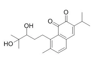
| Clin Cancer Res. 2005 May 1;11(9):3455-64. | | Antimetastatic effect of salvicine on human breast cancer MDA-MB-435 orthotopic xenograft is closely related to Rho-dependent pathway.[Pubmed: 15867248] | Salvicine is a novel DNA topoisomerase II inhibitor with potent anticancer activity. In present study, the effect of Salvicine against metastasis is evaluated using human breast carcinoma orthotopic metastasis model and its mechanism is further investigated both in animal and cellular levels.
METHODS AND RESULTS:
The MDA-MB-435 orthotopic xenograft model was applied to detect the antimetastatic effect of Salvicine. Potential target candidates were detected and analyzed by microarray technology. Candidates were verified and explored by reverse transcription-PCR and Western blot. Salvicine activities on stress fiber formation, invasion, and membrane translocation were further investigated by immunofluorescence, invasion, and ultracentrifugal assays. Salvicine significantly reduced the lung metastatic foci of MDA-MB-435 orthotopic xenograft, without affecting primary tumor growth obviously. A comparison of gene expression profiles of primary tumors and lung metastatic focus between Salvicine-treated and untreated groups using the CLOTECH Atlas human Cancer 1.2 cDNA microarray revealed that genes involved in tumor metastasis, particularly those closely related to cell adhesion and motility, were obviously down-regulated, including fibronectin, integrin alpha3, integrin beta3, integrin beta5, FAK, paxillin, and RhoC. Furthermore, Salvicine significantly down-regulated RhoC at both mRNA and protein levels, greatly inhibited stress fiber formation and invasiveness of MDA-MB-435 cells, and markedly blocked translocation of both RhoA and RhoC from cytosol to membrane.
CONCLUSIONS:
The unique antimetastatic action of Salvicine, particularly its specific modulation of cell motility in vivo and in vitro, is closely related to Rho-dependent signaling pathway. | | Biochem Biophys Res Commun. 2004 Oct 15;323(2):660-7. | | Telomerase inhibition is a specific early event in salvicine-treated human lung adenocarcinoma A549 cells.[Pubmed: 15369801 ] | The telomere and telomerase have been suggested as targets for anticancer drug discovery. However, the mechanisms by which conventional anticancer drugs affect these targets are currently unclear. The novel topoisomerase II inhibitor, Salvicine, suppresses telomerase activity in leukemia HL-60 cells. To further determine whether this activity of Salvicine is specific to the hematological tumor and distinct from those of other conventional anticancer agents, we studied its effects on telomere and telomerase in a solid lung carcinoma cell line, A549.
METHODS AND RESULTS:
Differences in telomerase inhibition and telomere erosion were observed between salvcine and other anticancer agents. All anticancer agents (except adriamycin) induced shortening of the telomere, which was identified independent of replication, but only Salvicine inhibited telomerase activity in A549 cells under conditions of high concentration and short-term exposure. At the low concentration and long-term exposure mode, all the tested anticancer agents shortened the telomere and inhibited telomerase activity in the same cell line. Notably, Salvicine inhibited telomerase activity more severely than the other agents examined. Moreover, the compound inhibited telomerase activity in A549 cells indirectly in a concentration- and time-dependent manner. Salvicine did not affect the expression of hTERT, hTP1, and hTR mRNA in A549 cells following 4 h of exposure. Okadaic acid protected telomerase from inhibition by Salvicine.
CONCLUSIONS:
These results indicate specificity of Salvicine and diversity of anticancer agents in the mechanism of interference with telomerase and the telomere system. Our data should be helpful for designing the study in the development of agents acting on telomere and/or telomerase. | | Mol Cancer Res. 2008 Feb;6(2):194-204. | | Salvicine inactivates beta 1 integrin and inhibits adhesion of MDA-MB-435 cells to fibronectin via reactive oxygen species signaling.[Pubmed: 18314480] | Integrin-mediated adhesion to the extracellular matrix plays a fundamental role in tumor metastasis. Salvicine, a novel diterpenoid quinone compound identified as a nonintercalative topoisomerase II poison, possesses a broad range of antitumor and antimetastatic activity.
METHODS AND RESULTS:
Here, the mechanism underlying the antimetastatic capacity of Salvicine was investigated by exploring the effect of Salvicine on integrin-mediated cell adhesion. Salvicine inhibited the adhesion of human breast cancer MDA-MB-435 cells to fibronectin and collagen without affecting nonspecific adhesion to poly-l-lysine. The fibronectin-dependent formation of focal adhesions and actin stress fibers was also inhibited by Salvicine, leading to a rounded cell morphology. Furthermore, Salvicine down-regulated beta(1) integrin ligand affinity, clustering and signaling via dephosphorylation of focal adhesion kinase and paxillin. Conversely, Salvicine induced extracellular signal-regulated kinase (ERK) and p38 mitogen-activated protein kinase (MAPK) phosphorylation. The effect of Salvicine on beta(1) integrin function and cell adhesion was reversed by U0126 and SB203580, inhibitors of MAPK/ERK kinase 1/2 and p38 MAPK, respectively. Salvicine also induced the production of reactive oxygen species (ROS) that was reversed by ROS scavenger N-acetyl-l-cysteine. N-acetyl-l-cysteine additionally reversed the Salvicine-induced activation of ERK and p38 MAPK, thereby maintaining functional beta(1) integrin activity and restoring cell adhesion and spreading.
CONCLUSIONS:
Together, this study reveals that Salvicine activates ERK and p38 MAPK by triggering the generation of ROS, which in turn inhibits beta(1) integrin ligand affinity. These findings contribute to a better understanding of the antimetastatic activity of Salvicine and shed new light on the complex roles of ROS and downstream signaling molecules, particularly p38 MAPK, in the regulation of integrin function and cell adhesion. |
|


 Cell. 2018 Jan 11;172(1-2):249-261.e12. doi: 10.1016/j.cell.2017.12.019.IF=36.216(2019)
 Cell Metab. 2020 Mar 3;31(3):534-548.e5. doi: 10.1016/j.cmet.2020.01.002.IF=22.415(2019)
 Mol Cell. 2017 Nov 16;68(4):673-685.e6. doi: 10.1016/j.molcel.2017.10.022.IF=14.548(2019)