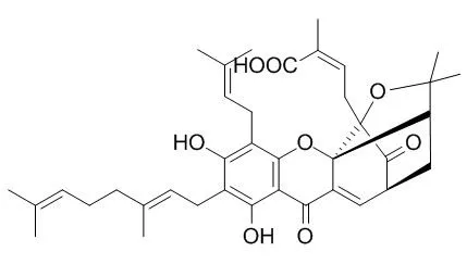
| Kinase Assay: |
| Zhongguo Zhong Yao Za Zhi. 2014 May;39(9):1666-9. | | Study of gambogenic acid-induced apoptosis of melanoma B16 cells through PI3K/Akt/mTOR signaling pathways.[Pubmed: 25095381] | To discuss the mechanism of Gambogenic acid (GNA) in inducing the apoptosis of melanoma B16 cells.
METHODS AND RESULTS:
The inhibitory effect of Gambogenic acid on the proliferation of B16 cells was measured by the methyl thiazolyl tetrazolium (MTT) assay. The effect of Gambogenic acid on B16 cells was detected by the Hoechst 33258 staining. The transmission electron microscopy was used to observe the ultra-structure changes of B16 cells. The changes in PI3K, p-PI3K, Akt, p-Akt, p-mTOR, PTEN proteins were detected by the Western blotting to discuss the molecular mechanism of Gambogenic acid in inducing the apoptosis of B16 cells. Gambogenic acid showed a significant inhibitory effect in the growth and proliferation of melanoma B16 cells. The cell viability remarkably decreased with the increase of Gambogenic acid concentration and the extension of the action time. The results of the Hoechst 33258 staining showed that cells processed with Gambogenic acid demonstrated apparent apoptotic characteristics. Under the transmission electron microscope, B16 cells, after being treated with Gambogenic acid, showed obvious morphological changes of apoptosis. The Western blot showed a time-dependent reduction in the p-PI3K and p-Akt protein expressions, with no change in p-PI3K and p-Akt protein expression quantities. The p-mTOR protein expression decreased with the extension of time, where as the PTEN protein expression showed a time-dependent increase.
CONCLUSIONS:
Gambogenic acid could inhibit the proliferation of melanoma B16 cells and induce their apoptosis within certain time and concentration ranges. Its mechanism in inducing the cell apoptosis may be related to PI3K/Akt/mTOR signaling pathways. | | PLoS One. 2014 Jan 10;9(1):e83604. | | Gambogenic acid kills lung cancer cells through aberrant autophagy.[Pubmed: 24427275] | Lung cancer is one of the most common types of cancer and causes 1.38 million deaths annually, as of 2008 worldwide. Identifying natural anti-lung cancer agents has become very important. Gambogenic acid (GNA) is one of the active compounds of Gamboge, a traditional medicine that was used as a drastic purgative, emetic, or vermifuge for treating tapeworm.
Recently, increasing evidence has indicated that Gambogenic acid exerts promising anti-tumor effects; however, the underlying mechanism remains unclear.
METHODS AND RESULTS:
In the present paper, we found that Gambogenic acid could induce the formation of vacuoles, which was linked with autophagy in A549 and HeLa cells. Further studies revealed that Gambogenic acid triggers the initiation of autophagy based on the results of MDC staining, AO staining, accumulation of LC3 II, activation of Beclin 1 and phosphorylation of P70S6K. However, degradation of p62 was disrupted and free GFP could not be released in Gambogenic acid treated cells, which indicated a block in the autophagy flux. Further studies demonstrated that Gambogenic acid blocks the fusion between autophagosomes and lysosomes by inhibiting acidification in lysosomes. This dysfunctional autophagy plays a pro-death role in Gambogenic acid-treated cells by activating p53, Bax and cleaved caspase-3 while decreasing Bcl-2. Beclin 1 knockdown greatly decreased Gambogenic acid-induced cell death and the effects on p53, Bax, cleaved caspase-3 and Bcl-2. Similar results were obtained using a xenograft model.
CONCLUSIONS:
Our findings show, for the first time, that Gambogenic acid can cause aberrant autophagy to induce cell death and may suggest the potential application of Gambogenic acid as a tool or viable drug in anticancer therapies. |
|
| Cell Research: |
| Zhong Yao Cai. 2014 Jan;37(1):95-9. | | Gambogenic acid induces mitochondria-dependent apoptosis in human gastric carcinoma cell line.[Pubmed: 25090714] | To study the effects of Gambogenic acid (GNA) on the growth of human gastric carcinoma cell line MGC-803 and its underlying mechanisms.
METHODS AND RESULTS:
MTT assay was used to measure the cell viability. Apoptosis, mitochondrial membrane potential (MMP), reactive oxygen species (ROS) were detected using flow cytometry method. Among them, Annexin V-FITC/PI double staining was employed in the analysis of apoptosis, Rh123 in analyzing MMP and H2DCFDA in analyzing ROS formation. P53 expression was confirmed by Western blot. 4.0 micromol/L Gambogenic acid inhibited MGC-803 cells growth in a time dependent manner from 24 to 48 h. At the concentration range from 1.0 to 12.0 micromol/L, the inhibitory effect was in a concentration dependent manner. After treatment with 4.0 micromol/L Gambogenic acid for 48 h, apoptosis was obviously observed as assayed by Annexin V-FITC/PI staining. Importantly, MMP was decreased and ROS formation was increased following Gambogenic acid treatment. Additionally, P53 expression was up-regulated following 4.0 micromol/ L Gambogenic acid treatment in a time dependent manner.
CONCLUSIONS:
Gambogenic acid induces mitochondria-dependent apoptosis and increases P53 expression in human gastric carcinoma cell line. |
|


 Cell. 2018 Jan 11;172(1-2):249-261.e12. doi: 10.1016/j.cell.2017.12.019.IF=36.216(2019)
 Cell Metab. 2020 Mar 3;31(3):534-548.e5. doi: 10.1016/j.cmet.2020.01.002.IF=22.415(2019)
 Mol Cell. 2017 Nov 16;68(4):673-685.e6. doi: 10.1016/j.molcel.2017.10.022.IF=14.548(2019)